2.17 Autoimmune Skin Diseases – Large Animals
Learning Objectives
- Learn that autoimmune skin diseases are rare in large animals.
- Know that pemphigus foliaceus has been reported in horses and goats and bullous pemphigoid in horses and pigs.
- Learn that discoid lupus erythematosus and systemic lupus erythematosus have been reported in horses.
- Learn how to recognize these diseases clinically.
- Learn that the diagnosis should be based on a characteristic history, clinical signs, cytological exam, histopathological findings, direct and indirect immunofluorescence, and antinuclear antibody (ANA) if suspecting of systemic lupus erythematosus.
- Know that the control of autoimmune skin diseases is often difficult requiring high doses of systemic glucocorticoids, with or without other potent immunomodulating drugs. Additionally, therapy usually is maintained for prolonged periods if not for life. Client education is essential for successfully managing these conditions.
- Remember to monitor for potential side effects when using the drugs used to treat autoimmune skin diseases. The same protocols described for dogs and cats described in previous sections can be used.
-
General Considerations
- Autoimmune skin diseases are rare in large animals.
- Pemphigus foliaceus has been reported in horses and goats.
- Bullous pemphigoid has been reported in horses and Yucatan mini pigs.
- Discoid lupus erythematosus and systemic lupus erythematosus have been reported in horses.
Important Facts
- Autoimmune skin diseases are rare in large animals.
- Most cases have been reported in horses and less frequently in goats, sheep, and pigs
-
Pathogenesis
- The pathogenesis of each of the autoimmune skin diseases mentioned below is the same described for dogs and cats in a previous section of Volume 2. Remember that drugs can trigger some of these diseases; therefore, a thorough drug history is important.
-
Clinical Signs
- Pemphigus foliaceus:
- It is a rare disease described in horses and goats.
- It is the most common form of pemphigus and the most common autoimmune skin disease of horses.
- There is no apparent age, breed, or sex predilection.
- Anecdotal reports indicate that triggering factors such as drug administration, systemic diseases, or stress precedes the onset of pemphigus foliaceus in horses.
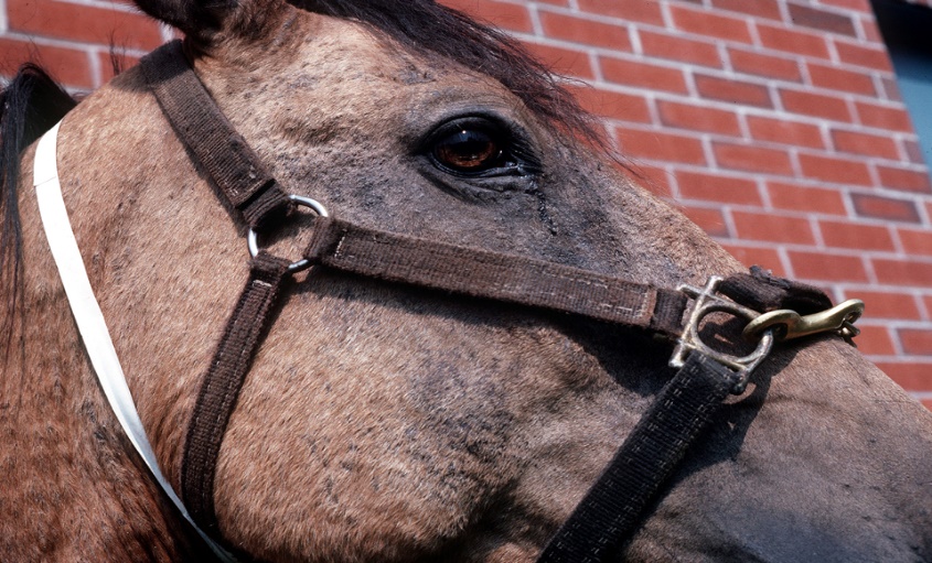
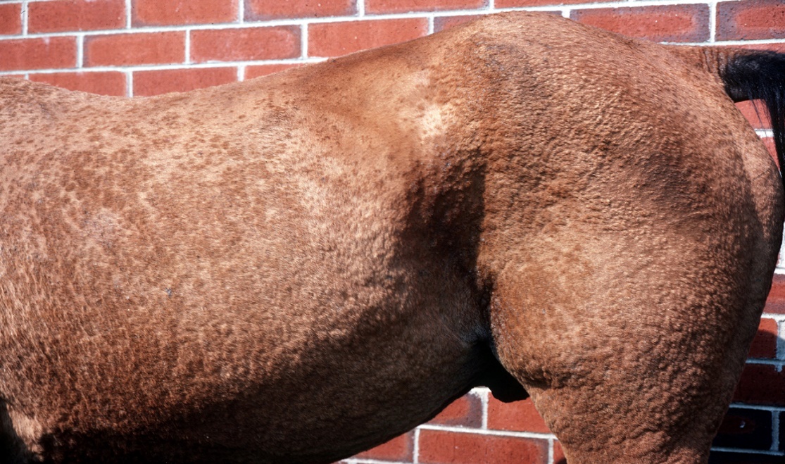
- In horses, skin lesions usually begin on the face or limbs including the coronary band, and become widespread. More than 50% of the cases have varying degrees of edema of the distal extremities and ventrum and it may be the first sign in some cases. Limb edema may occur in the absence of surface lesions.
- Pemphigus foliaceus:
-
-
- In goats, skin lesions usually begin on the ventrum and perineum, then become widespread with very severe lesions on the face and pinnae.
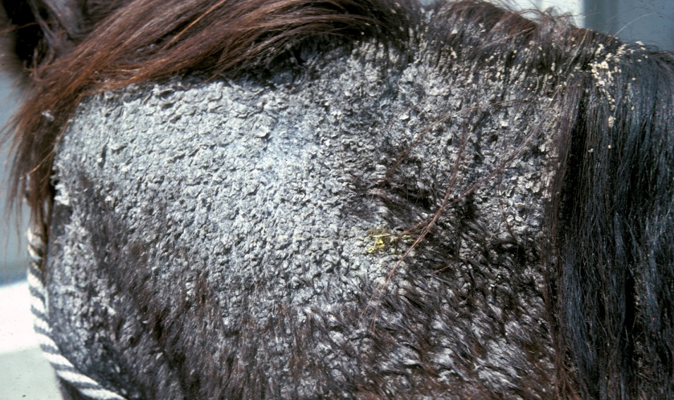
- The primary lesions are subcorneal or intracorneal pustules. However, these lesions are transient and the clinician usually sees tufted hairs associated with crusts, scales, oozing, annular erosions bordered by epidermal collarets. Alopecia and erythema are also often seen.
-
-
-
- Some horses show localized or more often generalized extensive exfoliative dermatitis.
-
-
-
- Pruritus and pain are variable.
- A juvenile or neonatal form with a better prognosis compared to adult horses has been reported.
- Over 50% of the horses and occasionally some goats have concurrent signs of systemic illness (i.e. depression, lethargy, weight loss, poor appetite, and pyrexia).
-
Important Facts
- Pemphigus foliaceus is the most common form of pemphigus and the most common autoimmune skin disease of horses.
- In horses, skin lesions usually start on the face or limbs and then become generalized.
- Edema of the extremities and ventrum can be the initial sign in some horses.
- In goats, skin lesions start on the ventrum and perineum then become generalized affecting also the face and pinnae.
- Similar to small animals, primary lesions are pustules. These lesions are transient and most of the time the clinician will see tufted hairs associated with crusts, scales, oozing, and annular erosions.
- Over 50% of the horses and occasionally some goats have concurrent signs of systemic illness (i.e. depression, lethargy, weight loss, poor appetite, and pyrexia).
-
- Bullous pemphigoid:
- Bullous pemphigoid has been rarely reported in horses of all ages (5-14 years) and breeds.
- Signs usually develop slowly over several weeks,
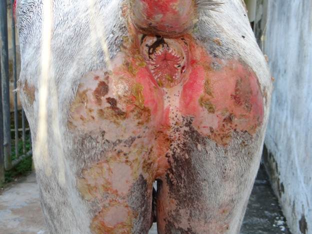
- The primary lesions are vesicles or bullae. These lesions will rupture very easily leaving crusts, deep erosions and more often ulcerations.
- Bullous pemphigoid:
-
-
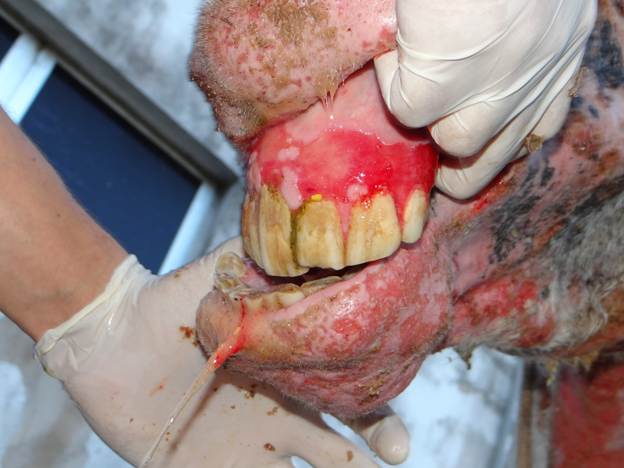
- The areas affected are the mucocutaneous junctions (lips, vulva, anus, eyelids), oral cavity, nasal mucosa and intertriginous areas (most commonly axillary and groin regions).
-
-
-
- Pain and pruritus are variable.
- Severely affected horses may be anorectic, depressed, lose weight and febrile.
- It has also been reported in Yucatan minipigs, affecting mainly their dorsum.
-
Important Facts
- Bullous pemphigoid has been rarely reported in horses and Yucatan minipigs.
- The lesions are characterized by vesicles or bullae, which rupture easily resulting in crusts, deep erosions and/or ulcerations.
- Lesions are typically localized to the oral cavity, nasal mucosa, mucocutaneous junctions, and axillary and inguinal regions.
- Severely affected horses may be anorectic, depressed, febrile, and lose weight.
-
- Systemic Lupus Erythematosus:
- Systemic lupus erythematosus is a multisystemic autoimmune disorder that has been rarely recognized in horses.
- There are no reported age, breed, and sex predilections.
- The clinical signs are variable.
- Cutaneous changes have included lymphedema of the extremities, panniculitis, mucocutaneous ulceration, alopecia, generalized exfoliative dermatitis, leukoderma, and scaling of the face, neck, and trunk.
- Extracutaneous abnormalities have included polyarthritis, thrombocytopenia, peripheral lymphadenopathy, proteinuria, fever, depression, anorexia, and weight loss.
- Sun exposure aggravates the disease.
- Systemic Lupus Erythematosus:
Important Facts
- Systemic lupus erythematosus is a multisystemic autoimmune disease rarely recognized in horses, with variable clinical signs.
- Cutaneous changes have included lymphedema of the extremities, panniculitis, mucocutaneous ulceration, alopecia, generalized exfoliative dermatitis, leukoderma, and scaling of the face, neck, and trunk.
- Other non-cutaneous signs have included polyarthritis, thrombocytopenia, peripheral lymphadenopathy, proteinuria, fever, depression, anorexia, and weight loss.
-
- Discoid Lupus Erythematosus:
- Discoid lupus erythematosus has been rarely recognized in horses.
- There are no apparent breed and sex predilections.
- Affected horses range from1 to 14 years of age.
- It is a relatively benign skin disease without systemic involvement. Affected horses are otherwise healthy.
- Patchy erythema, alopecia, scaling, and crusting are usually localized to the face, periocular areas, nostrils, ears, and neck. The perineal, perianal and genital areas may be affected.
- Leukoderma and leukotrichia may be seen within lesions.
- Pruritus and pain are usually mild to absent.
- Sun exposure aggravates the disease.
- Discoid Lupus Erythematosus:
Important Facts
- Discoid lupus erythematosus has been rarely recognized in horses.
- Patchy erythema, alopecia, scaling, and crusting are usually localized to the face, periocular areas, nostrils, ears, and neck. The perineal, perianal, and genital areas may be affected.
- Sun exposure aggravates the disease; therefore, it should be avoided.
- Affected horses are otherwise healthy.
-
Diagnosis
- The information obtained from a thorough patient’s history and clinical signs is important to obtain a diagnosis or a sensible list of differential diagnoses.
- Nikolsky sign is usually positive in bullous pemphigoid and epidermolysis bullosa.
- Cytologic exam:
- Pemphigus foliaceus:
- Direct smears of the exudate collected from intact pustules, from recent erosions, and/or from the exudate underneath a crust, reveal numerous acantholytic keratinocytes, numerous non-degenerate neutrophils or eosinophils, and no intracellular bacteria (however, they can be seen if secondary infection is present).
- Occasional acantholytic keratinocytes can be present in any suppurative condition, but when seen in clusters or large numbers in several microscopic fields they are strongly suggestive of pemphigus. In horses, dermatophytic infections caused by Trichophyton spp. have been associated with profound acantholysis; therefore, it is recommended to perform special stain of skin biopsy and fungal culture to rule out dermatophytosis in any case suggestive of pemphigus foliaceus in horses.
- Bullous pemphigoid, epidermolysis bullosa, discoid lupus erythematosus, and systemic lupus erythematosus:
- Cytologic exam does not demonstrate acantholytic keratinocytes.
- Pemphigus foliaceus:
- Clinical Pathology:
- More than 50% of the horses with pemphigus foliaceus manifest some combination of non- regenerative anemia, neutrophilia, hypoalbuminemia, and elevations of serum globulins.
- Skin biopsy: formalin fixed samples.
- Pemphigus foliaceus:
- Intact pustules are the ideal sample. Biopsy a recent crust and erosion if pustules are not present. Remember to not surgically prep the skin before biopsying and to biopsy through the crust (i.e. include the crust in your sample since acantholytic cells may be imbedded in the crust).
- Multiple biopsies taking samples from different stages of disease development will greatly increase the chances of demonstrating diagnostic histopathological changes.
- Pemphigus foliaceus is characterized by subcorneal or intragranular acantholysis with resultant cleft and pustule formation. Hair follicles may also be affected.
- Neutrophils and/or eosinophils may predominate within the pustule. These inflammatory cells will accompany the acantholytic keratinocytes.
- Bullous pemphigoid:
- Numbers a and b under “Pemphigus foliaceus” are also applicable for bullous pemphigoid.
- Histologically lesions are characterized by subepidermal vacuolar alteration and subepidermal cleft and vesicle formation.
- Dermal inflammatory infiltrates are usually mild to moderate and perivascular.
- Acantholytic keratinocytes are not present.
- Discoid lupus erythematosus and systemic lupus erythematosus:
- Skin biopsy reveals hydropic and/or lichenoid interface dermatitis.
- Multiple biopsies taking samples from different stages of disease development will greatly increase the chances of demonstrating diagnostic histopathological changes.
- Acantholytic keratinocytes are not present.
- Pemphigus foliaceus:
- Direct immunofluorescence (skin biopsy samples are fixed in Michel’s solution) or immunohistochemistry (skin biopsy samples are fixed in formalin):
- Pemphigus foliaceus:
- Diffuse intercellular deposition of immunoglobulin (and occasionally complement) within the epidermis.
- Direct immunofluorescence has been reported to be positive in equine dermatophilosis as well. Results must be interpreted in conjunction with the patient’s history, clinical signs, cytological exam, and histopathology.
- Bullous pemphigoid:
- Linear deposition of immunoglobulin and usually complement at the basement membrane zone of affected skin or mucosae.
- Discoid lupus erythematosus and systemic lupus erythematosus:
- Deposition of immunoglobulin or complement, or both, at the basement membrane zone.
- Pemphigus foliaceus:
- Indirect immunofluorescence (serum is collected for this test):
- It is usually not requested routinely to diagnose autoimmune skin diseases because the results are often negative. Additionally, this test is not widely available.
- Clinical pathology:
- Perform a CBC, chemistry profile, urinalysis and joint fluid analysis if you suspect systemic lupus erythematosus or if the animal is systemically ill.
- Antinuclear antibody test (ANA):
- It is generally positive for systemic lupus erythematosus and negative for the other autoimmune skin diseases.
Important Facts
- The information obtained from a detailed patient’s history and clinical signs are very important diagnostic tests.
- Cytologic examination of the exudate collected from an intact pustule or vesicle or from a recent crust or erosion will reveal large numbers of acantholytic keratinocytes, neutrophils, and eosinophils in cases of pemphigus foliaceus.
- More than 50% of the horses with pemphigus foliaceus manifest some combination of non-regenerative anemia, neutrophilia, hypoalbuminemia, and elevations of serum globulins.
- Bullous pemphigoid, epidermolysis bullosa, discoid lupus erythematosus, and systemic lupus erythematosus do not demonstrate acantholytic keratinocytes.
- Histopathology is a very important diagnostic test for all autoimmune skin diseases. Samples are preserved in formalin for this test.
- Try to biopsy primary lesions (i.e pustules, vesicles, or bullae). If not possible, biopsy a recent crusty lesion or a recent erosive lesion.
- Multiple biopsies taking samples from different stages of disease development will greatly increase the chances of demonstrating diagnostic histopathological changes.
- Direct immunofluorescence and immunohistochemistry tests can be used to support a presumptive diagnosis. Remember! False positive and false negative results can occur with these tests. Try to sample vesicles, pustules, or bullae for these tests.
- Preserve the skin biopsy sample in Michel’s solution for direct immunofluorescence and in formalin for immunohistochemistry.
- Indirect immunofluorescence is not routinely requested to diagnose autoimmune skin diseases because false negative results occur often.
- Serum should be collected for indirect immunofluorescence because it is evaluating the presence of circulating autoantibodies.
- Perform a CBC, chemistry profile, urinalysis, and joint fluid analysis if you are suspecting of systemic lupus erythematosus.
- Antinuclear antibody test (ANA) should be positive in systemic lupus erythematosus and negative in the other autoimmune skin diseases.
-
Treatment: Pemphigus foliaceus, bullous pemphigoid, discoid lupus erythematosus, and systemic lupus erythematosus.
-
- Treatment of autoimmune skin diseases is often difficult requiring high doses of systemic glucocorticoids with or without other potent immunomodulating drugs.
- Additionally, therapy is often maintained for prolonged periods if not for life because autoimmune skin diseases cannot be cured.
- Client education is essential to obtain a successful outcome.
- High dose of systemic glucocorticoid is the initial treatment of choice:
- Prednisolone: 1.0-2.0 mg/kg given orally twice daily.
- Dexamethasone: 0.2-0.4 mg/kg given orally once daily.
- Disease control is usually achieved within 7 to 10 days, at which point alternate-morning therapy is begun and the dose is slowly reduced to the lowest possible dosage.
- When systemic glucocorticoids are unsatisfactory, the addition or substitution of other immunomodulating drug may be required for an adequate disease control.
- Gold salts (aurothiomalate, Myochrisine®) or azathioprine can be added to the glucocorticoid regimen or used in place of glucocorticoids. Remember that these drugs have a long lag phase (i.e. azathioprine 4 to 6 weeks; gold salts 6 to 12 weeks). Pentoxifylline, which improves tissue oxygenation and has an anti-inflammatory effect can also be used as adjunctive therapy in horse cases. This is a very safe drug.
- Azathioprine: 2.0 mg/kg q 24h until remission of signs then every 48 hr.
- Gold salts: 1 mg/kg intramuscularly per week until remission, then once a month.
- Pentoxifylline: 10-12 mg/kg q 12h.
- Remember to monitor for potential side effects when using azathioprine and gold salts drugs (same tests and protocol described for dogs and cats – refer to previous section of these notes).
- Sun avoidance is an important part of the treatment regimen of autoimmune skin disorders.
Important Facts
- Treatment of autoimmune skin diseases is often difficult requiring high doses of systemic glucocorticoids, with or without other potent immunomodulating drugs.
- Additionally, therapy usually is maintained for prolonged periods, if not for life, because autoimmune skin disease are not curable.
- Client education is essential for successfully managing these conditions.
- High dose of systemic glucocorticoid is the initial treatment of choice of many dermatologists.
- Control of the dermatosis is usually achieved within 7 to 10 days, at which point alternate-morning therapy is begun and the dose is slowly reduced to the lowest possible dosage.
- When systemic glucocorticoids are unsatisfactory, the addition or substitution of other immunomodulating drug may be required for an adequate disease control.
- Remember to monitor for potential side effects when using these drugs.
- Sun avoidance is an important part of the treatment regimen of autoimmune skin diseases.
References
Knottenbelt DC. Chapter11: Immune-mediated/allergic diseases. In: Pascoe’s Principles & Practice of Equine Dermatology. 2009. W.B. Saunders, p. 253-297.
Medeiros, Gildenor X, Riet‐Correa F. Epidermolysis bullosa in animals: a review. Vet Dermatol 2015; 26(1): 3-e2.
Scott, DW. Color atlas of farm animal dermatology 2008. John Wiley & Sons.
Scott DW. Immunologic Disease. In: Large Animal Dermatology 1985. W.B. Saunders, Philadelphia, p307-317.
Scott DW. Miller WH. Chapter 9: Immune-mediated disorders. In: Equine Dermatology 2003. W.B. Saunders, St. Louis, p. 475-547.
Vandenabeele, SIJ et al. Pemphigus foliaceus in the horse: a retrospective study of 20 cases. Vet Dermatol 2004; 15(6): 381-388.
