3.13 Canine Zinc Responsive Dermatosis
-
General Considerations
- Zinc-responsive dermatosis is a rare chronic keratinization disorder that responds to zinc supplementation.
- Zinc is an important component of many metalloenzymes that participate in regulating the metabolism especially of rapidly dividing cells such as the keratinocytes.
- Two clinical syndromes are recognized.
- Syndrome I occurs in dogs that are on balanced diets.
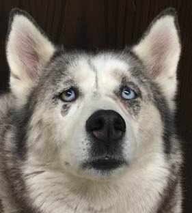
- Siberian huskies and Alaskan malamutes are the breeds most frequently representing Syndrome I. However, Syndrome I has also been reported in Bull terriers and other breeds albeit less frequently.
- The pathomechanism is not well understood but decreased intestinal absorption of zinc has been identified in some Alaskan malamutes.
- The age of onset has been reported to range from 6 months to 10 years, with 41% of dogs developing lesions before 2 years old.
- This condition may be precipitated by stress, estrus, and severe gastrointestinal disorders affecting absorption.
- Diets high in phytate (i.e. important plant component that stores phosphorus) may also precipitate the disorder by binding to zinc in the gastrointestinal tract and interfering with its absorption.
- Syndrome II affects mainly rapidly growing puppies or young adults on zinc-deficient diets or dietary regimens that involve high concentrations of calcium, iron, and copper, which compete with zinc binding sites in the gastrointestinal tract and interfere with its absorption. This syndrome is also seen in dogs being fed diets high in phytate. Again, phytate binds to zinc and affects its absorption.
- Breeds reported with Syndrome II include Great Dane, Doberman pincher, beagle, German shorthair pointer, Labrador retriever, and Rhodesian ridgeback.
- Puppies or young adults of any breed may be affected if fed zinc-deficient diets or diets rich in components that interfere with zinc absorption.
- Severe forms of zinc-related dermatosis associated with zinc deficiency and a poor to guarded prognosis have been reported in bull terrier puppies and in a litter of Pharaoh hound dogs.
- The disease in bull terrier puppies is called “lethal acrodermatitis”. The condition is associated with inhibition of absorption and utilization of zinc and is inherited as an autosomal recessive trait.
Important Facts
- Zinc-responsive dermatosis is a rare chronic keratinization disorder that responds to zinc supplementation.
- Syndrome I occurs in dogs that are on balanced diets and is more common in the Siberian husky and Alaskan malamute dogs.
- Abnormal absorption of zinc has been reported in some Alaskan malamute dogs.
- Forty percent of dogs develop clinical signs before 2 years old.
- Diets high in calcium and phytate may precipitate the disorder by impeding zinc absorption in the gastrointestinal tract.
- Syndrome II affects mainly rapidly growing puppies or young adult dogs on zinc-deficient diets or regimens that involve high concentrations of minerals (especially calcium, iron, and cooper) and diets high in phytate.
- Syndrome II may occur in any breed fed diets deficient in zinc or containing components that impede zinc absorption.
- Severe forms of zinc-related dermatosis have been reported in bull terrier puppies and in a litter of Pharaoh hound dogs.
-
Clinical Signs
- In Syndrome I, clinical signs usually develop in young adults, 1 to 3 years of age, while in Syndrome II clinical signs occur in puppies prior to 1 year of age.
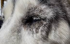
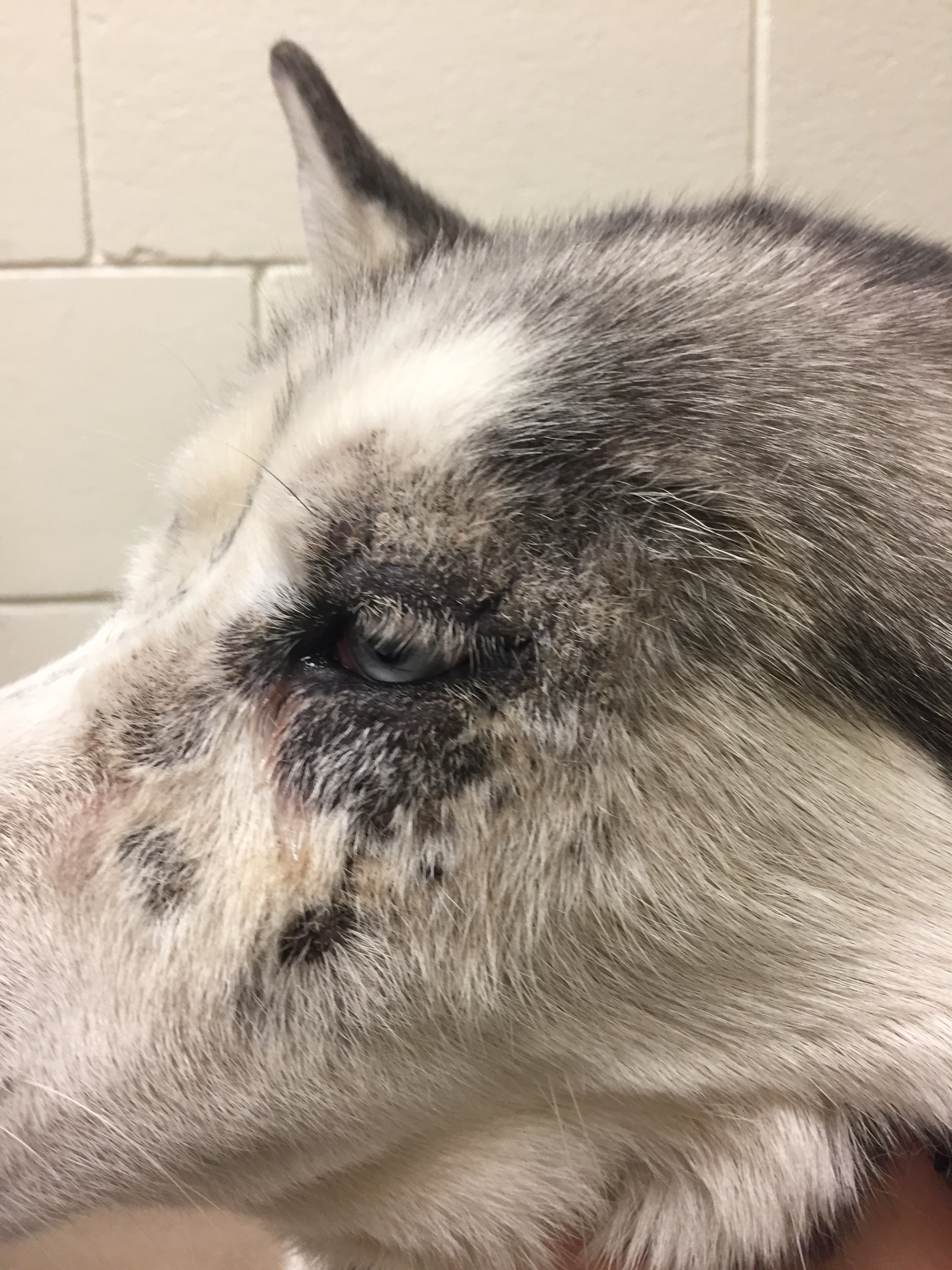
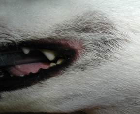
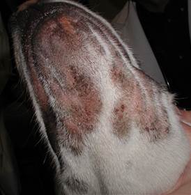
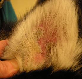
- Erythema, partial to complete alopecia, tightly adhered scales, and crusts are the predominant lesions.
-
- Distribution primarily involves the face including the periorbital region, muzzle, perioral area (commissures and lip margins), chin, and ears. The prepuce, scrotum, and vulva may also be affected.
-
- Heavy crusts or tightly adhered scales are also observed on pressure points, especially elbows and hocks.
- The footpads can become hyperkeratotic and onychomalacia may be seen.
- Hyperpigmentation of affected areas may be seen in chronic lesions.
-
- The hair coat may be dry and dull.
- Secondary bacterial infection and Malassezia and/or bacterial overgrowth are common and may contribute to the severity of the lesions and pruritus.
- A significant proportion of affected dogs exhibit pruritus, which may precede the development of cutaneous lesions.
Important Facts
- In Syndrome I, clinical signs usually develop in young adults 1 to 3 years of age being fed a balanced diet, while in Syndrome II clinical signs occur in puppies or young adults being fed a diet deficient in zinc or rich in components (e.g. calcium, iron, copper, and phytate) that impede zinc absorption.
- Erythema, alopecia, tightly adhered scales, and crusts are the predominant lesions.
- Lesions can be localized to one or more of the following sites: face, muzzle, periorbital and perioral regions, chin, ears, prepuce and pressure points, especially elbows and hocks.
- The footpads are often hyperkeratotic.
- Secondary bacterial infection and Malassezia and/or bacterial overgrowth are common and may aggravate preexisting lesions.
- A significant proportion of affected dogs exhibit pruritus, which may precede the development of cutaneous lesions.
-
Diagnosis
- A tentative diagnosis can be made based on the dog breed, age of disease onset, and clinical signs including lesion characteristics and distribution.
- Concentration of zinc in hair and serum of affected dogs is variable; which question the accuracy of these tests.
- Skin biopsy is needed to confirm a presumptive clinical diagnosis.
- Histopathology characterized by severe diffuse epidermal and follicular parakeratotic hyperkeratosis, epidermal hyperplasia, and dermal inflammation are findings supportive of zinc responsive dermatosis if the history and clinical signs are compatible.
- Differential diagnoses include demodicosis, dermatophytosis, Malassezia dermatitis, pemphigus foliaceus, generic dog food dermatitis, superficial necrolytic dermatitis, systemic lupus erythematosus, and mucocutaneous pyoderma.
- Skin scrapings and cutaneous cytology should be performed on all suspect cases.
- Response to therapy with zinc supplements supports the diagnosis.
Important Facts
- A tentative diagnosis can be made based on the breed, age of disease onset, lesion distribution, and appearance of gross lesions.
- Measurements of zinc concentrations in hair and serum are variable and unreliable.
- Skin biopsy is required to confirm a presumptive clinical diagnosis.
- Response to zinc supplementation supports the diagnosiss.
-
Treatment
- Oral zinc supplementation along with proper dietary correction when indicated, usually results in satisfactory control of clinical signs within 4 to 8 weeks.
- In some cases, resolution of signs can only be noticed after 3 to 7 months of therapy.
- Several forms of zinc supplement have been used.
- Zinc sulfate, zinc gluconate, and zinc methionine have been recommended.
- The dose of zinc sulfate is 10 mg/kg orally every 24 hrs.
- The dose of zinc gluconate and methionine, which are organic compounds containing zinc, should be based on their content of elemental zinc.
- In general, it is more useful to base dosages on the amount of elemental zinc because the zinc content in the various supplements may differ considerably.
- An oral dose of 2-3 mg of elemental zinc per kilogram of body weight every 24 hrs is the recommended initial dose.
- If vomiting occurs, the zinc supplement can be crushed and mixed with food. This will improve absorption and reduce gastrointestinal irritation.
- Zinc sulfate, zinc gluconate, and zinc methionine have been recommended.
- Treatment duration:
- Life-long therapy is necessary for dogs classified in the Syndrome I, as lesions generally recur within 2 to 8 weeks following discontinuation of zinc supplementation.
- In Syndrome II, oral zinc supplement may be given only for a few weeks to help restore zinc levels.
- All potential dietary imbalances should be corrected by feeding good quality balanced diets.
- In dogs presenting Syndrome II, dietary correction alone may resolve the skin lesions in 2-6 weeks.
- Phytase may be added to the diet as it enhances the bioavailability of zinc by hydrolyzing phytates present in foods.
- An alternative intravenous zinc therapy has been used in severe cases or cases refractory to oral supplementation, such as in bull terriers and Pharaoh dogs.
- Sterile zinc sulfate solutions (10 to 15 mg/kg diluted at 1:1 ratio with saline) are administered intravenously weekly for 4 weeks.
- Maintenance injections may be needed at intervals of 1 to 6 months to prevent relapses.
- Other treatments include:
- Systemic glucocorticoids at anti-inflammatory doses. They can significantly improve response to zinc supplementation and speed resolution of lesions. The favorable effects of glucocorticoids may result from their anti-inflammatory action in the skin and/or from improved gastrointestinal absorption of zinc.
- Essential fatty acids (EFA) supplementation may be beneficial. It has been documented that EFA deficiency impairs zinc absorption; therefore, EFA supplementation may enhance zinc absorption. In both syndromes, fasting and post-prandial concentrations of serum triglycerides have been shown to be significantly lower compared with normal dogs.
- Symptomatic therapy with warm water soaks (5-10 minutes) and antiseborrheic shampoos are indicated to loosen and remove the excessive tightly adhered scaling and crusting. Petrolatum and ointment-based anti-inflammatory and antimicrobial topical therapies may be used.
- Affected female dogs may improve after ovariohysterectomy as estrus may trigger the disease in predisposed dogs.
Important Facts
- Zinc supplementation usually results in satisfactory control of clinical signs within 4 to 8 weeks.
- Many dogs will need concurrent oral glucocorticoids as they improve zinc absorption, reduce skin inflammation, and the associated pruritus.
- Zinc sulfate, zinc gluconate, or zinc methionine has been recommended for the life of the animals with the Syndrome I.
- In dogs with Syndrome II, dietary correction alone may resolve the skin lesions in 2-6 weeks.
- In all cases, dietary imbalances should be corrected by feeding good quality balanced diets.
References
Kwochka KW: Primary Keratinization Disorders of Dogs. In: Griffin CE, Kwochka KW, MacDonald JM (eds). Current Veterinary Dermatology. St Louis, Mosby Year Book, 1993; p 176-190.
Kwochka KW: Overview of normal keratinization and cutaneous scaling disorders of dogs. In: Griffin CE, Kwochka KW, MacDonald JM (eds). Current Veterinary Dermatology. St Louis, Mosby Year Book, 1993; p 167-175.
Miller, Griffin and Campbell. Chapter 14. Keratinization defects. In: Muller & Kirk’s Small Animal Dermatology. 7th ed., W.B. Saunders, Missouri, 2013; p 630-646.
Power HT, Ihrke PJ. Synthetic retinoids in veterinary dermatology. Vet Clin North Am: Small Anim Pract, Philadelphia, WB Saunders, 1990; p 1525.
White SD et al. Zinc‐responsive dermatosis in dogs: 41 cases and literature review. Vet Dermatol 2001; 12(2): 101-109.
