2.9 Exfoliative Cutaneous Lupus Erythematosus
-
General Considerations
- Exfoliative cutaneous lupus erythematosus is a rare severe and disseminated form of cutaneous lupus erythematosus.
- It is reported to be hereditary and transmitted in an autosomal recessive manner in German shorthaired pointer dogs.
- ECLE is typically associated with systemic signs.
- It appears to have an autosomal recessive mode of inheritance.
- The pathogenesis of exfoliative cutaneous lupus erythematosus in dogs is not well known and specific antinuclear autoantibodies have not been identified in dogs. However, it is believed to be similar to the pathogenesis of other lupus diseases in dogs.
Important Facts
- Exfoliative cutaneous lupus erythematosus is a severe skin disease associated with systemic signs.
- It affects mostly young German shorthaired pointer dogs and an autosomal recessive mode of inheritance has been reported in this breed.
-
Clinical Signs
- Signalment: the information is based on a comprehensive review on canine cutaneous lupus erythematosus.
- Exfoliative cutaneous lupus erythematosus occurs predominantly in German shorthair pointer dogs. However, this disease has also been reported in several Magyar viszla dogs from Western Europe. Interestingly, viszlas and German shorthair pointers share a common ancestral.
- The disease was reported to occur 1.4 times more often in female than male dogs.
- The reported median age of disease onset is 8 months (range: 7 weeks to 3.5 years).
- Lesions: the information is based on a large case series of 25 dogs.
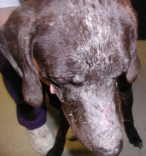
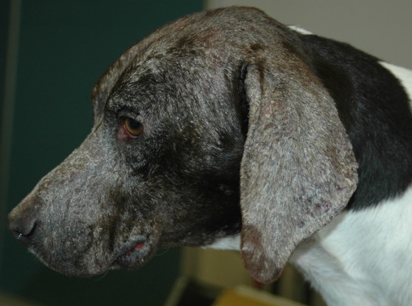
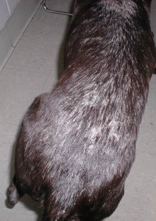
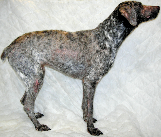
- Lesions usually start as scaling (100% of the reported cases), alopecia (76%) and follicular casts (28%). The disease can progress to develop erosions, ulcers and crusts (25%), and it is generalized in most cases.
- Signalment: the information is based on a comprehensive review on canine cutaneous lupus erythematosus.
-
- Distribution:
- Lesions usually start on the face (i.e. muzzle, forehead, pinnae), and dorsum and progress to affect the ventral trunk and limbs.
- Distribution:
-
- Pruritus may be present.
- Generalized peripheral lymphadenopathy and intermittent pyrexia may occur.
- Episodes of arthralgia manifested as stiffness and/or shifting leg lameness, hunched stance, and reluctance to move, can develop in many dogs as the disease progresses.
-
- Hematologic abnormalities include anemia, lymphopenia, and thrombocytopenia.
- Progressive infertility (i.e. low sperm count and abnormal estrous cycles) has been reported.
Important Facts
- Skin lesions are characterized by exfoliation, alopecia, erosions, ulcers, and crusts on face, trunk, and extremities.
- Affected animals are usually sick with lymphadenopathy, pyrexia, and arthralgia.
-
Diagnosis
- Differential diagnoses:
- Sebaceous adenitis, pemphigus foliaceus, and systemic lupus erythematosus. However, these diseases typically affect much older dogs.
- Characteristic history, signalment, and clinical signs are important parts of the diagnostic equation.
- Cytology:
- No specific findings and no acantholytic cells. This test is important to investigate the presence of secondary infection.
- Histopathology:
- Hyperkeratosis and lymphocytic interface dermatitis extending to the infundibulum of the hair follicles. Apoptosis of the basal keratinocytes, with or without lymphocytic satellitosis, is common but the apoptosis can also be seen in the upper epidermal layers. Involvement of sebaceous glands is common. There may be features similar to sebaceous adenitis, with sebaceous glands being absent in half of all sections evaluated in a case series of 25 dogs with 16% of these dogs having no sebaceous glands. .
- Select scaling, alopecic, and eroded sites to biopsy.
- Direct immunofluorescence or immunohistochemistry:
- In a case series of 25 dogs, deposition of IgG immunoglobulin in the epidermal and follicular basement membrane was reported in 100% and 41% of the test dogs, respectively.
- History, clinical signs, and histopathologic findings are the most valuable diagnostic tools.
- Preserve samples in Michel’s solution for immunofluorescence and formalin for immunohistochemistry.
- Indirect immunofluorescence:
- In a case series of 25 dogs, circulating anti-follicular and anti-sebaceous gland IgG antibodies were demonstrated by indirect immunostaining in 57% of the tested dogs, with no evidence of autoantibodies against the dermal-epidermal junction.
- Antinuclear antibody (ANA) test:
- Negative
- CBC may show mild anemia and fluctuating thrombocytopenia. Serum chemistry profile may show hyperglobulinemia but in general, the abnormalities are not consistent. Joint fluid analysis in dogs with intermittent arthralgia, failed to identify any abnormality.
- Differential diagnoses:
Important Facts
- Diagnosis should be based on a suggestive signalment (i.e. young German short-haired pointer dogs) and history (progressive skin disease associated with pain), characteristic clinical signs (i.e. exfoliative skin disease and arthralgia), and histopathological findings (i.e. hyperkeratosis and lymphocytic interface dermatitis).
- ANA is negative.
-
Treatment
- The prognosis is generally guarded and worsens with aging and progression of arthralgia.
- Many dogs are euthanized due to either failure to respond to therapy or complications associated with immunomodulatory therapy.
- One or a combination of the following drugs: (See below “Therapy for Autoimmune Diseases” for dose and specific on treatment regimen).
- Glucocorticoids.
- Azathioprine.
- Mycophenolate mofetil.
- Oclacitinib (Apoquel®) – Complete remission was achieved in a dog treated with oclacitinib.
- Hydroxychloroquine sulfate.
- Cyclosporine.
References
Mauldin EA, Morris DO, Brown DC, Casal ML. Exfoliative cutaneous lupus erythematosus in German shorthaired pointer dogs: disease development, progression and evaluation of three immunomodulatory drugs (ciclosporin, hydroxychloroquine, and adalimumab) in a controlled environment. Vet Dermatol 2010; 21(4):373-382.
Medleau L, Hnilica KA. Chapter 8. Autoimmune and immune-mediated skin disorders. In: Small Animal Dermatology: A color Atlas and Therapeutic Guide 2006. 2nd ed. W.B. Saunders, Missouri, 189-227.
Miller, Griffin and Campbell. Chapter 9. Autoimmune and immune-mediated dermatoses. In: Muller & Kirk’s Small Animal Dermatology 2013. 7th ed., W.B. Saunders, Missouri; 432-462.
Olivry T. Auto-immune skin disease in animals: time to reclassify and review after 40 years. BMC Vet Res 2018; https://doi.org/10.1186/s12917-018-1477-1.
Olivry T, Chan LS. Autoimmune blistering dermatoses in domestic animals. Clin Dermatol 2001; 19(6):750-760.
Olivry T., Jackson HA. Diagnosing new autoimmune blistering skin diseases of dogs and cats. Clin Tech Small Anim Pract 2001; 16: 225-229.
Olivry T, Linder KE, Banovic F. Cutaneous lupus erythematosus in dogs: a comprehensive review. BMC Vet Res 2018; https://doi.org/10.1186/s12917-018-1446-8
