2.9 Urticaria – Small and Large Animals
Learning Objectives
- Know that urticaria can be precipitated by immunologic and nonimmunologic factors.
- Identify the types of hypersensitivity involved in the immunologically-induced urticaria.
- Know the causes and various factors that precipitate and/or aggravate urticaria.
- Describe the clinical manifestation of urticaria.
- Describe how you would diagnose and treat cases of urticaria.
-
General Considerations
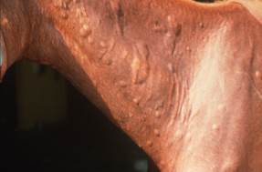
- Urticaria is a disease characterized by the sudden development of wheals (hives) and angioedema or both.
- A wheal is a transient elevated lesion that results from dermal edema and inflammatory cell infiltration.
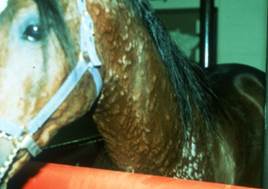
- Angioedema is characterized by a sudden swelling of the deep dermis and subcutis. The swelling is pronounced and the affected skin becomes erythematous. The body sites often affected include the lower extremities and head. Wheals may or may not be present.
- Urticaria is typically associated with a type I (immediate, IgE mediated) hypersensitivity but other immune mechanisms can be involved and it can also be nonimmunologic in nature.
- Urticaria can occur in any animal species but it is most commonly seen in horses and dogs.
Important Facts
- Urticaria is a disease characterized by the sudden development of wheals and angioedema or both.
- It can be triggered by an immunologic mediated (typically IgE mediated) or nonimmunologic mediated response to various stimuli.
- Urticaria can occur in any animal species but it is most commonly seen is horses and dogs.
-
Cause and Pathogenesis
- In urticaria, activated mast cells and basophils release histamine and other mediators (e.g. platelet-activating factor, cytokines etc.), which cause vasodilatation, plasma extravasation, sensory nerve activation, and recruitment of inflammatory cells. These events participate in lesion formation.
- The activating response can be immunologically mediated or nonimmunologically mediated.
- The immunological reactions are mainly immunoglobulin (Ig) E driven (type I hypersensitivity), although type II and type III hypersensitivity (immune complexes) may also be involved. Examples of triggers include allergens, infectious agents (e.g. bacteria, parasites) or autoantibodies.
- Nonimmunologic factors that may precipitate or intensify urticarial reactions are cold, heat, sunlight, exercise, stress, genetic influences and various drugs and chemicals (e.g. aspirin, narcotics, foods, food additives).
- Factors reported to have caused urticaria are various:
- Drug: antibiotics, phenothiazines (thiabendazole, acepromazine), local anesthetics; nonsteroidal anti-inflammatory drugs (phenylbutazone), etc.
- Food – fish, grain, hay, chemical additives, etc.
- Environmental: pollen, mold, grain dust, etc.
- Envenomation: snake or insect bites/stings.
- Plant: nettle, buttercup, etc.
- Vaccine.
- In dogs, the triggers of urticaria that are supported by studies or case reports are vaccines, anesthetics, foods, venoms, drugs, transfusions, plants and radiocontrast media. Some rare causes such as heat, exercise, sunlight, dermatographism, estrous, intestinal parasites, allergen-specific immunotherapy and intradermal testing are only anecdotally reported in veterinary textbooks.
Important Facts
- Urticaria can result from immunologic and nonimmunologic stimuli.
- Immunologic cases are typically associated with a type I (IgE mediated) hypersensitivity reaction.
- Nonimmunologic factors that may precipitate or intensify urticarial reaction are cold, heat, sunlight, exercise, stress, genetic influences and various drugs and chemicals (e.g. aspirin, narcotics, foods, food additives).
-
Clinical Signs
- Clinical signs are similar in all species and will be discussed together.
- No age predilection has been reported for urticaria. In dogs, short coated breeds such as boxers and pit bulls appear to be predisposed. However, it is possible that urticaria is easier to visualize in these breeds.
- There may be a female predisposition.
- A study demonstrated that dogs with a history of atopic dermatitis seem to be at increased risk of developing urticaria or anaphylaxis regardless of cause.
-
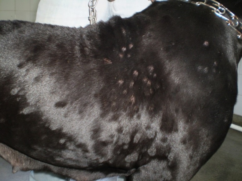
- Reactions in all species are characterized by localized or generalized wheals, which usually do not exhibit serum leakage or hemorrhage.
- Lesions appear suddenly and typically regress within a day, but occurrence of new lesions may persist over a longer period if the culprit is not identified.
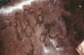
- Urticarial lesions may occasionally assume bizarre patterns (serpiginous, linear, arciform, annular, papular), especially in horses. Hair may appear raised in these areas.
-
- Pruritus may or may not be present.
Important Facts
- No age, sex or breed predilection has been reported.
- Reactions are characterized by localized or generalized wheals, which usually do not exhibit serum leakage or hemorrhage.
- Urticarial lesions may occasionally assume bizarre patterns (serpiginous, linear, arciform, annular, papular). Hair may appear raised in these areas.
- Pruritus may or may not be present.
-
Diagnosis
- Diagnosis is made based on a detailed history and typical clinical signs.
- Biopsy can be performed to confirm the diagnosis and rule out other conditions that may present similarly such as, erythema multiforme, vasculitis, bacterial folliculitis, and mastocytosis.
- The difficult part is finding the triggering cause.
- It is important to take a thorough history including information about exposure to drugs, vaccines, dewormers, supplements, and feeds. Remember that in some cases, urticaria is triggered by cold, heat, exercise, physical trauma, or pressure.
- If pruritus is present, a food elimination trial should be considered in small animals (8 weeks) and in large animals (4 weeks). If food allergy is ruled out, atopic dermatitis is a reasonable diagnosis and intradermal testing for environmental allergens (e.g. molds, pollens of trees, grasses, weeds, house dust mites etc.) may be indicated in some cases with the goal of trying allergen-specific immunotherapy.
Important Facts
- Diagnosis is based on a thorough history and characteristic clinical signs.
- It may be difficult or impossible to find the trigger.
-
Treatment
- Elimination and avoidance of culprit, if possible.
- Symptomatic treatment with systemic glucocorticoids.
- Large animals – 0.1 mg/kg dexamethasone, IV or IM, or 1 mg/kg of prednisolone, IV or IM.
- Small animals – prednisone or prednisolone at 2 mg/kg PO, IM or IV.
- In horses with chronic urticaria, pentoxifylline (8–10 mg/kg PO twice to three times daily) should also be tried. This drug has been shown to stabilize mast cells in other species.
- Antihistamines are used to prevent future reactions or for chronic urticaria and are only minimally helpful for pruritus.
- Epinephrine may be lifesaving in severe angioedematous or anaphylactic reactions.
- Large animals: 3 to 5 ml of a 1:100 solution, SQ or IM.
- Small animals: 0.1 to 0.5 ml of a 1:1000 solution SQ or IM.
- Drug–related urticaria usually respond poorly to glucocorticoids and can persist for several months after the last administration of the drug (though most do not).
Important Facts
- Elimination and avoidance of known etiologic factors, if possible.
- Symptomatic therapy with glucocorticoids and antihistamines.
- Epinephrine may be lifesaving in severe angioedematous or anaphylactic reactions.
References
Fadok VA. Update on equine allergies. Vet Clin Equine 2013; 29: 541–550.
Hinden S, Klukowska‐Rötzler J, Janda J et al. Characterization of the inflammatory infiltrate and cytokine expression in the skin of horses with recurrent urticaria. Vet Derm 2012; 23: 503.
Marcella R, White S, Fadok VA, et al. Equine allergic skin diseases: Clinical consensus guidelines of the World Association for Veterinary Dermatology. Vet Derm 2023; 34: 175-208.
Marsella R. Equine allergy therapy. Vet Clin North Am Equine Pract 2013; 29: 551-557.
Miller, Griffin, Campbell. Muller and Kirk’s Small Animal Dermatology. 7th Edition. In: Hypersensitivity Disorders. Elsevier Health Sciences. 2013 p 363-364.
Rostaher A, Hofer‐Inteeworn N, Kümmerle‐Fraune C et al. Triggers, risk factors and clinico‐pathological features of urticaria in dogs–a prospective observational study of 24 cases. Vet Derm 2017; 28: 38.
Scott, DW. Large Animal Dermatology. In: Immunologic Diseases. W.B. Saunders, Philadelphia, PA, 1988, p 292 – 294.
