4.9 Subcutaneous Mycosis – Eumycotic Mycetoma – Small and Large Animals
Learning Objectives
- Know that mycetoma lesions are characterized by tumefaction, draining tracts and grains mixed in the exudate.
- Learn that a mycetoma can be caused by fungus (i.e. eumycotic) or bacteria (i.e. actinomycotic – Actinomyces spp., Nocardia spp.).
- Know that the animals develop a cutaneous to subcutaneous nodule at the site of entry, which drains a serous-sanguineous or purulent exudate containing grains.
- Know that the grains in eumycotic mycetomas consist of broad septate branching hyphae and can be seen on cytology of aspirates or direct smears and in biopsy tissues. A granulomatous to pyogranulomatous inflammation is associated with grains.
- Also know that the fungal elements grow on Sabouraud’s dextrose agar. The best samples for culture are either tissue grains or tissue fragments.
- Learn how to manage eumycotic mycetoma.
-
General Considerations
- The cardinal signs of eumycotic mycetoma are:
- Tumefaction.
- Draining tracts (not always present).
- Grains (granules) in the exudate from draining tracts.
- The grains are composed of necrotic material and aggregates of fungal elements. They can be black or white depending on the fungus causing the infection.
- The disease is rare in the U.S. and Europe. It is more commonly seen in Central and South America, India, and southern Asia.
- The cardinal signs of eumycotic mycetoma are:
Important Facts
- Eumycotic mycetoma is caused by fungal organisms.
- The cardinal signs of mycetomas include tumefaction, draining tracts (not always present) and grains mixed in the exudate from draining tracts.
- The disease is rare in the U.S. and Europe and is more common in Central and South America, India, and southern Asia.
-
Etiology
- The agents are ubiquitous soil saprophytes.
- White-grain fungi:
- Acremonium sp.
- Pseudoallescheria boydii (most common in the U.S.).
- White-grain mycetomas typically present as chronic intra-abdominal or body wall granulomas that develop months to years after a contaminated wound or surgical site, especially after dehiscence.
- Black-grain fungi:
- Curvularia spp.
- Exophiala jeanselmei
- Leptosphaeria spp.
- Pyrenochaeta spp.
- Staphylotrichum spp.
- Torula spp.
- Helminthosporium speciferum.
- Allescheria boydii.
- Maduralla spp.
- Bracycladium spicaterum.
- White-grain fungi:
- The agents are ubiquitous soil saprophytes.
Important Facts
- The grains can be black or white depending on the fungus causing the infection.
- Pseudoallescheria boydii is the most common etiologic agent in the U.S.
-
Pathogenesis
- The disease is typically caused by wound contamination with the saprophytic fungi that live in the soil.
-
Species
- Mycetomas have been reported in dogs, cats, and horses.
-
Clinical Signs
- Most cutaneous infections are caused by black-grain fungal organisms.
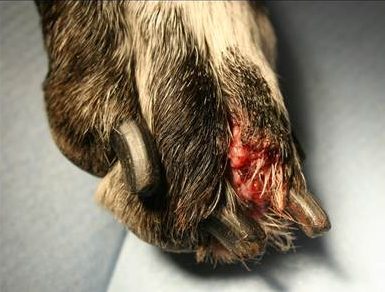
- The limbs and face are the sites most commonly affected and the lesions are usually solitary.
- Subcutaneous masses are often painful and develop at site of fungal entry. Draining tracts develop and exude a serous, purulent, or hemorrhagic discharge. Grains present in the discharge vary in size, shape and color depending on the fungus involved.
-
- Cycles of healing and active lesions may result to firm swelling and scarring.
- Extension of infection to the underlying muscle, joint and bone can occur in chronic infections.
- The disease can become disseminated.
- Differential diagnosis include:
- Other subcutaneous or deep fungal infection.
- Kerion dermatophyte lesion.
- Abscess.
- Foreign body skin reaction.
- Mycobacterial infection (opportunistic mycobacterial).
- Sterile nodular panniculitis.
- Sterile pyogranuloma.
- Neoplasia.
Important Facts
- Lesions are usually solitary and occur most commonly on the limbs and face.
- Subcutaneous masses drain a serous, purulent or hemorrhagic exudate, which contains grains of different sizes, shapes and colors depending on the fungus involved.
- Cutaneous lesions are typically associated with black-grain fungus.
- The disease can become systemic.
-
Diagnosis
- A detailed history and characteristic clinical signs are important parts of the diagnostic equation.
- Cytology: Cytological examination of lesion aspirates and direct smears of exudate from draining tracts show a pyogranulomatous inflammation with rare fungal elements. Fungal organisms can be seen by squashing the grains. For detail on the cytological features of the fungal organisms, refer to the review paper by Dehghanpir 2023.
- Skin biopsy: Histopathological findings include a pyogranulomatous to granulomatous inflammation with the presence of fungal elements associated with grains within the inflammatory reaction. To learn about the histopathologic features of the grains, refer to the review article by Hoffmann et al, 2023.
- Fungal culture: The fungal organisms grow in Sabouraud’s dextrose agar. Grains and tissue are the preferred samples for culture.
- PCR test identifies the fungal species.
Important Facts
- The diagnosis is based on a detailed history, characteristic clinical signs, and results of one or more of the following tests: cytology of lesion aspirates and direct smears of exudate, histopathology and fungal culture.
- PCR test identifies the fungal species.
-
Treatment: Refer to the paper by Dedeaux and the Blastomycosis chapter for information on dose protocols of antifungals
- Wide surgical excision is the treatment of choice. However, depending on the size and location of the lesion, it may not be possible.
- Itraconazole, posaconazole, voriconazole or terbinafine can be used after surgery when wide margins cannot be achieved. Ideally, antifungal chemotherapy should be based on in vitro susceptibility testing of the isolate.
- It is also important to know that medical therapy likely will not work by itself because adequate concentration of the antifungal drug will not reach the granules where the fungal organism lives.
- Treatment must be continued for 2 to 3 months after clinical cure.
Important Facts
- Wide surgical excision is the treatment of choice but it may not be possible in all cases.
- Any attempt at antifungal chemotherapy should be ideally based on susceptibility testing of the isolate.
- Antifungal chemotherapy likely will not work as monotherapy because adequate concentration of the antifungal drug will not reach the granules where the fungus resides.
- Treat for 2 to 3 months after clinical cure.
References
Dedeaux A, Grooters A, Wakamatsu-Utsuki N et al. Opportunistic fungal infections in small animals. J Am Anim Hosp Assoc 2018; 54: 327–337. DOI 10.5326/JAAHA-MS-6768
Dehghanpir SD. Cytomorphology of deep mycoses in dogs and cats. Vet Clin Small Anim 2023; 53: 155–173.
Greene CE. Infectious Diseases of the Dog and Cat. 4th ed. St. Louis, Missouri, Elsevier, Saunders, 2012.
Hoffmann AR, Ramos MG, Walker RT et al. Hyphae, pseudohyphae, yeasts, spherules, spores, and more: A review on the morphology and pathology of fungal and oomycete infections in the skin of domestic animals. Vet Pathol 2023; doi.org/10.1177/03009858231173715
Lichon V and Khachemoune A. Mycetoma A Review. Am J Clin Dermatol 2006; 7: 315-321.
McEntee M. Eumycotic mycetoma: review and report of a cutaneous lesion caused by Pseudallescheria bodyii in a horse. J Am Vet Med Assoc 1987; 191: 1459-1461.
Miller WH, Griffin CE, Campbell KL. Muller & Kirk’s Small Animal Dermatology. 7th ed. St. Louis, Missouri, Elsevier, Mosby, 2013.
Rippon JW. Medical Mycology. Philadelphia, WB Saunders, 1988.
Scott DW. Large Animal Dermatology. Philadelphia, WB Saunders, 1988.
Verma P and Jha A. Mycetoma: reviewing a neglected disease. Clin Exp Dermatol 2019; 44:123-129.
