1.6 Subcutaneous Abscesses – Large Animals
Learning Objectives
- What is (are) the most common etiologic agent(s) causing abscesses in swine, horses, cattle, goats and sheep?
- What predisposes an animal to develop a subcutaneous abscess?
- How do you manage an abscess?
- What causes caseous lymphadenitis in goats and sheep?
- How is the disease characterized clinically?
- How do you diagnose caseous lymphadenitis?
- What is the best way to manage this condition?
- What are important preventative measures you must adopt?
-
General Considerations
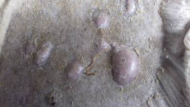
- Cutaneous abscesses are circumscribed accumulations of pus that typically extend to the subcutaneous tissue.
- Cutaneous abscesses are common in large animals.
- They usually develop as a result of bacterial contamination of cutaneous wounds.
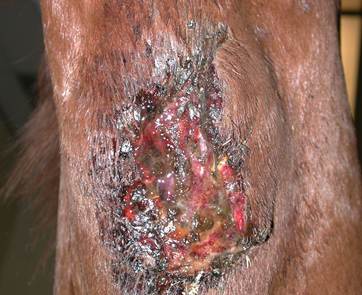
- In general, abscesses are characterized initially by firm, often painful and warm nodules that progress to having a soft, fluctuant texture. Typically, they drain in the later stages.
- Caseous lymphadenitis will also be discussed in this section, but separately.
-
Etiology and Clinical Signs
- Swine:
- Most commonly caused by Corynebacterium pyogenes and B-hemolytic streptococci. Less commonly caused by Fusobacterium necrophorum, Staphylococcus hyicus, Bacteroides and Clostridium species.
- Skin abscesses occur more commonly on the shoulders, neck, ears, flank, tail, and feet, which are sites more susceptible to trauma.
- Mortality rate in young animals can be up to 30%.
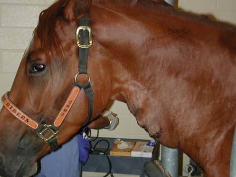
- Horses:
- Typically caused by Corynebacterium pseudotuberculosis, Clostridium species, Streptococcus equi and Staphylococcus species.
- Common sites include the hoof, cheek, chest, shoulder and rump (often associated with injections).
- Swine:
-
-
- Streptococcus equi causes strangles and abscesses are formed in the submandibular and especially parotid lymph nodes. Systemic dissemination of the infection in severe cases will results in the formation of cutaneous abscesses in various parts of the body.
- Goats:
- Typically caused by Corynebacterium pseudotuberculosis, Arcanobacterium pyogenes, Escherichia coli, Staphylococcus spp. and Streptococcus spp.
- Abscesses often develop as a result of trauma to the skin and oral mucosa.
- Common causes include self-biting of the cheek due to manual restraint of the head in a way that the cheek mucosa is forced between the molar teeth or the use of tight halters; skin trauma from grass awns or thorny vegetation; bite wounds from predators or other goats; dehorning; vaccination, ear tagging etc.
- Sheep:
- Usually caused by Corynebacterium pseudotuberculosis and Corynebacterium pyogenes.
- Sporadic abscesses in sheep are typically caused by skin trauma such as, shearing, vaccination, ear tagging, rough hay racks or milking stanchions, bite wounds from predators or other sheep in situations of overcrowding, and skin penetration of plant awns, thorns or other foreign bodies.
-
-
-
- The traumatized skin becomes then an open door for various bacterial sources such as, resident skin bacteria, soil and fecal bacteria, or oral bacteria.
- Bovine:
- Usually caused by Corynebacterium pyogenes.
-
-
Diagnosis
- History and clinical signs.
- Cytological examination of exudate.
- Bacterial culture and sensitivity.
-
Treatment
- Systemic antibiotic is contraindicated prior to lancing because it prevents the abscess from heading-up.
- Hot pack, bring abscess to a head, lance and flush with compounds such as povidone iodine or diluted chlorhexidine.
- Systemic antibiotic therapy based on culture and sensitivity results can be used if topical therapy is ineffective or multiple lesions and systemic signs are present.
-
Caseous Lymphadenitis
-
General Considerations:
- Caseous lymphadenitis is considered one of the most serious diseases of goats in the U.S.
- It is the leading cause of condemnation of sheep carcasses.
- It is caused by Corynebacterium pseudotuberculosis and it is the most common cause of abscess formation in sheep and goats.
- Any abscess localized to a lymph node of sheep or goats should be presumed to be caused by Corynebacterium pseudotuberculosis until proven otherwise.
-
Etiology and Pathogenesis:
- Corynebacterium pseudotuberculosis (also called Corynebacterium ovis), a gram-positive, facultative intracellular bacterium.
- Organisms enter the body by ingestion or through skin wounds (especially shearing).
- May occur 6 months after initial infection.
- The organism can live on fomites for about 55 days, soil for about 8 months and water for about 60 hours.
-
Clinical Signs:
- Abscesses of lymph nodes and subcutaneous tissues (nodules and draining tracts in the skin).
- Abscesses contain thick, pasty, curd-like yellowish, greenish or tan material.
- Abscesses may open and persistently drain or become encapsulated.
- Most common sites are the head (submandibular and parotid lymph nodes) and neck (prescapular lymph node) but it can occur anywhere on the body.
- Abscesses may involve internal lymph nodes in the chest.
- Animals of any age may be affected.
- Usually, the disease is brought into a herd by an infected new animal.
- The disease may progress to affect internal lymph nodes and organs.
-
Diagnosis:
- Differential diagnosis include impacted cud, salivary cyst, wattle cyst, neoplasia, and goiter. In addition, abscesses caused by other bacterial pathogens such as, Actinobacillus licheniformis, Arcanobacterium pyogenes and Staphylococcus aureus subsp. anaerobius should be considered as potential causes but the infection with these organisms is sporadic and does not affect various animals in a flock.
- History and physical findings.
- Cytology of purulent exudate collected from abscesses. Try to sample early lesions because no organisms may be seen in older lesions as they are in few numbers.
- Culture: organism takes several days to grow on blood agar. Select early lesions. Make sure to disinfect the skin before sampling because saprophytic bacteria may overshadow Corynebacterium pseudotuberculosis on culture medium.
- Antibody ELISA tests are available.
- PCR can be used for direct detection of Corynebacterium pseudotuberculosis in exudate from lesions or culture colonies. Blood samples are not useful to detect the bacterium via PCR.
-
Treatment:
- Isolation.
- Open abscess, drain and pack with iodine or antibiotic. Dispose of the purulent material carefully to prevent environmental contamination.
- Removing an intact abscess by surgical excision is the best treatment.
- Systemic antibiotics are often not efficacious because of the thick abscess capsule that impedes the antibiotic penetration.
-
Prevention:
- Isolate all new animals: examine monthly after quarantine period.
- Early recognition of abscesses.
- Infected animals should be isolated until the abscess is completely healed, or surgical intervention or euthanasia is performed.
- Chronically affected animals should be culled from flock or herd.
- Purchase replacement animals from farms with low abscess herd history.
- Decrease crowding in pens.
- Kids should be isolated from affected mothers and should not be fed their milk if they are being kept as replacements.
- Sheep- shear young animals before adults and disinfect shearing equipment.
- Vaccine does not provide absolute immunity but should be considered in endemic areas. They are more useful at restraining dissemination of an infection and will not eliminate an already established infection.
-
Important Facts
- Cutaneous abscesses are common in large animals.
- They usually develop because of bacterial contamination of cutaneous wounds.
- In general, abscesses are characterized by firm, often painful and warm nodules that progress to having a soft, fluctuant texture. Typically, they drain in the later stages.
- Treatment includes thorough drainage of the abscess with povidone iodine or diluted chlorhexidine. Systemic antibiotic is rarely needed and should be avoided.
- Corynebacterium pseudotuberculosis is the most common cause of abscesses in goats and sheep.
- The clinical disease is called caseous lymphadenitis.
- Caseous lymphadenitis is considered one of the most serious diseases of goats in the U.S. and is the leading cause of condemnation of sheep.
- Abscesses associated with this disease contain thick, pasty, curd-like yellowish/greenish material.
- Removing intact abscesses by surgical excision is the best treatment.
- Systemic antibiotics are not generally helpful and should be avoided.
- Animals with caseous lymphadenitis should be isolated until the abscess is completely healed, or surgical intervention or euthanasia can be performed.
References
Ayers, J.L.: Caseous Lymphadenitis. In: Howard, J.L. (ed.): Current Veterinary Therapy. Food Animal Practice. Philadelphia, W.B. Saunders Co., 1981, p 660.
Oreiby AF. Diagnosis of caseous lymphadenitis in shepp and goat. Small Anim Res 2015; 123:160-166.
Scott DW. Bacterial Diseases. In: Large Animal Dermatology. Philadelphia, PA: W.B. Saunders, 1988; 135-136; 150.
Scott DW, Smith MC, Manning TO. Caprine Dermatology. I. Normal Skin and Bacterial and Fungal Disorders. Comp Cont Ed 1984; 6:S 190.
Williamson LH. Caseous lymphadenitis in small ruminants. Vet Clin North Am Food Anim Pract 2001; 17:359-371.
Windsor PA and Bush RD. Caseous lymphadenitis: Present and near forgotten from persistent vaccination? Small Anim Res 2016; 142:6-10.
