5.1 Poxvirus Infections – Large Animals
Learning Objectives
- Know that poxviruses belong to a large family of DNA viruses that share group-specific nucleoprotein antigens.
- Know that poxvirus infections can be transmitted to humans. So, be careful when managing a suspected case! Transmission is by direct and indirect contact, and human-to-human transmission can occur. The most common zoonotic diseases are pseudocowpox, contagious viral pustular dermatitis and papular stomatitis.
- Know that lesions caused by poxviruses are very characteristic. Erythematous macules evolve to papules, which evolve to vesicles (not always present). Vesicles evolve to umbilicated pustules, which are characterized by a central depression and a raised and often erythematous edge. This typical lesion is called pock. The pustules rupture and form crusts. The lesions often heal with a scar.
- Know that the diagnosis is based on a detailed history and characteristic clinical signs and should be supported by one or more of the following tests: histopathology, electron microscopy, viral identification via serologic and immunofluorescence techniques and molecular tests.
- Remember! The disease is self-limited and it usually takes a few weeks to resolve. Treatment when required involves symptomatic and supportive care.
-
General Considerations
- Poxiviruses belong to the poxiviridae family of DNA viruses that share group-specific nucleoprotein antigens.
- The genera include Orthopoxvirus (cowpox and vaccinia), Capripoxvirus (sheep-pox, goatpox, and bovine lumpy skin disease), Suipoxvirus (swinepox), and Parapoxvirus (pseudocowpox, bovine papular stomatitis, and contagious viral pustular dermatitis).
- Horsepox virus, the virus of ovine viral ulcerative dermatosis, equine viral papular dermatitis, and molluscum contagiosum, remains unclassified.
- Humans can develop skin lesions from many of the poxviruses of large animals including vaccinia, cowpox, goatpox, pseudocowpox, bovine papular stomatitis, horsepox, and contagious viral pustular dermatitis.
-
- The poxviruses that are more easily transmitted to humans include viral pustular dermatitis, pseudocowpox, and bovine papular stomatitis.
- We will only discuss the diseases diagnosed in the United States.
Important Fact
- Poxviruses are DNA viruses that share group-specific nucleoproteins.
- The poxviruses that are more easily transmitted to humans include the agents of viral pustular dermatitis, pseudocowpox, and bovine papular stomatitis.
-
Pathogenesis
- Infection typically occurs via cutaneous viral inoculation or inhalation.
- Poxviruses commonly gain access to the systemic circulation via the lymphatic system, and rarely via the blood stream.
- A second viremia leads to the dissemination of the virus back to the skin and other organs.
Important Fact
- Infection typically occurs via cutaneous or respiratory routes.
- In most cases, the first viremia occurs via the lymphatic system, and rarely via the blood stream.
- A second viremia leads to the dissemination of the virus back to the skin and other organs.
-
Clinical Signs
- Cutaneous pox lesions have a characteristic clinical evolution:
- Lesions begin as erythematous macules, which evolve to papules and then vesicles.
- The vesicular stage is well developed in some pox infections but ephemeral or absent in others.
- Vesicles evolve into pustules with a depressed center and an erythematous and raised border. This lesion is known as pock.
- Yellowish to brownish crusts are formed after the pustules rupture.
- It is common for scars to form when lesions heal.
- Cutaneous pox lesions have a characteristic clinical evolution:
Important Fact
- Lesions begin as erythematous macules, which evolve to papules and then vesicles.
- Vesicles evolve into pock lesions characterized by pustules with a depressed center and raised, typically erythematous border.
- The pustule ruptures easily and crusts form as the exudate dries.
- Healed lesions often leave a scar.
-
- We will discuss the poxvirus infections diagnosed in the United States:
- Goatpox:
- Capripoxvirus causes goatpox infection of goats. The disease has been reported in Africa, Asia, parts of Europe, and the United States.
- There is no age predisposition; however, the disease seems to be more severe in young animals.
- Mortality is typically less than 5%; however, morbidity can be as high as 90%.
- Transmission of infection occurs by contact through abraded skin, inhalation, or mechanically by flies (Stomoxys calcitrans).
- The reported incubation period ranges from 5 to 17 days.
- At the start of the infection, the animals typically have pyrexia, anorexia, conjunctivitis, and rhinitis. About 2 to 3 days later, skin lesions develop on various parts of the body.
- Lesions can be limited to one body region such as, the muzzle and lips.
- Lesions typically resolve in 3 to 4 weeks.
- Humans in contact with infected goats can develop pox-like lesions on the hands and forearms.
- Swinepox:
- Swinepox has a worldwide distribution and can become endemic in areas of intensive swine production.
- It is caused by a suipoxvirus.
- The swine louse, Haematopinus suis, has been implicated as a vector in the spread of swine pox.
- Pigs less than 3-4 months of age are more commonly infected than adults are but it can affect all ages.
- Mortality is typically low but morbidity may be high.
- The disease is often associated with poor sanitation.
- The incubation period ranges from 2 to 6 days.
- Systemic illness is rarely present.
- Skin lesions follow the pock sequence and occur most commonly on the non-haired portions of the body.
- Secondary bacterial infection is common and may obscure the primary condition.
- Healing typically occurs within 2 to 4 weeks and lifelong immunity develops after recovery.
- Recovered animals serve as the reservoir of infection for the remainder of the herd.
- Vaccination is not usually practiced and control of the pig lice is the principal prophylactic measure attempted in most outbreaks.
- Humans in contact with affected pigs do not develop skin lesions.
- Pseudocowpox:
- Pseudocowpox infection occurs in cattle around the world.
- It is caused by a parapoxvirus.
- Minimal persistent immunity appears to occur after infection, thus cyclic surges of reinfection occur within a herd.
- Morbidity is very high.
- There is no age predilection, and milking cows and heifers appear to be most susceptible.
- Transmission most commonly occurs at milking time and is mechanical, with the potential for transmission from cow to calf by suckling.
- Difficulty in milking and an increased incidence of mastitis result in economic losses.
- The incubation period is about 6 days.
- Clinical signs are quite variable and affected animals do not develop systemic illness.
- Lesions are generally located on the lips, in the oral cavity, and/or on the muzzle in younger animals, and on the teats and udders in cows.
- The initial clinical signs in cows are erythema, focal edema and pain of affected teats. Pain is moderate and present only in the pre-scab stage. Vesicle formation is uncommon.
- After about 2 days, the lesion evolves to develop a “scab” that resembles a ring or a horseshoe. This occurs about 12-14 days later. The horseshoe crust is considered pathognomonic for pseudocowpox.
- Lesions typically resolve in about six weeks.
- Suggested measures to reduce discomfort when milking include (i) careful removal of the scabs, which should be burned to avoid contaminating the environment, (ii) application of an emollient ointment just before milking and thereafter, (iii) the use of an astringent, such as triple dye.
- An iodophor teat dip is recommended as the most effective control measure because it appears to exert some antiviral effect. An effort should be made to reduce teat trauma because infection is facilitated by discontinuity of the skin barrier.
- Pseudocowpox virus also cause skin lesions in humans; therefore, caution should be taking when handling affected cattle.
- Bovine Papular Stomatitis:
- It is caused by a parapoxvirus. It occurs in cattle all over the world.
- The disease is more common in animals younger than one year of age.
- Morbidity can be as low as 10% or reach 100% but rarely affected animals die of the disease.
- The incubation period is 3 to 5 days.
- The disease is usually mild and of limited economic importance.
- Pox-like lesions usually occur on the muzzle, nostrils and lips and, occasionally on the sides, abdomen, hind legs, scrotum, and prepuce. Teat lesions may develop in cows nursing infected calves.
- Other than the skin lesions, affected animals are healthy.
- It may take weeks to months before lesions resolve.
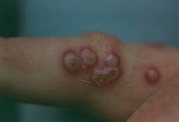
- It is a zoonotic disease and the virus causes skin lesions in humans, especially on the finger and hands. The lesions are known as “milker’s nodules”.
- Contagious Viral Pustular Dermatitis:
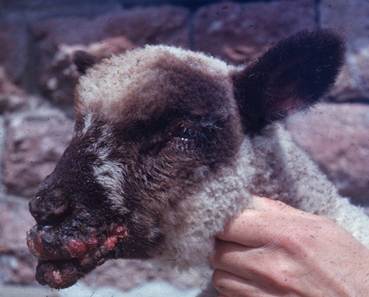
- Contagious viral pustular dermatitis is also known as contagious ecthyma, contagious pustular dermatitis, sore mouth, “scabby mouth” or ORF. It is a worldwide disease of sheep and goats and infrequently cattle.
- It is caused by an enveloped double stranded DNA virus, the orf virus, which belongs to the genus Parapoxvirus of the Poxviridae family.
- The economic loss can be significant due to failure to suck or graze, mastitis, abandonment, and related death. The importance of orf infection has recently increased due to the emergence of this virus in new territories, the occurrence of re-infection of previously infected animals, as well as interspecies infection.
- Morbidity may be as high as 90%, but mortality is typically 1% or less.
- The transmission within a herd is carried out through direct contact between animals during confrontation or suckling.
- The disease tends to be most common during the lambing and kidding seasons.
- Lambs and kids of three to six months of age appear to be more susceptible. It also occurs after an animal enters a feedlot.
- Skin lesions follow a typical pock progression.
- Lesions are quite proliferative and usually occur on the lips, muzzle, nostrils, around the mouth and eyelids. The disease may also result in genital, udder and foot lesions. In severe forms of disease, additional lesions can be observed in the oral mucosa.
- Goatpox:
- We will discuss the poxvirus infections diagnosed in the United States:
-
-
-
- Lesions may occur on the teats and udders of nursing ewes and does as well as the teats and udders of cattle.
- Healing typically occurs within few weeks. Scabs usually dry up and fall off in 1 to 4 weeks.
- Virus loses infectivity after 30 to 60 days of hot weather but it can survive over the winter.
- Post-recovery immunity usually lasts at least 1 year.
- No protective antibodies are passed in the colostrum, making the newborn of immune dams still susceptible.
- It is a zoonotic disease. Humans (farmers, butchers, sheep and goats shearers and veterinarians) can be infected by direct contact with sick animals. The preferential location of the lesions is the hand.
- Vaccination:
- If endemic, all previously unexposed need to be vaccinated.
- Vaccine is a live virus vaccine made from infected scabs suspended in glycerol saline.
- The vaccine is applied to scarified skin to produce lesions (usually inside of the rear leg, ear or under the tail).
- Three weeks are needed for complete immunity.
- Immunity is not passed in colostrum.
- Next year’s kid cohort will be infected unless annual vaccination of herd is continued.
- Vaccinated animals should not go to shows until all scabs have fallen off.
- Vaccine can cause lesions in humans – wear gloves.
- Ovine Viral Ulcerative Dermatosis:
- Ovine viral ulcerative dermatosis is a worldwide infectious disease of goats and sheep. It is also known as lip and leg ulceration, infectious balanoposthitis, ulcerative vulvitis, and ovine venereal disease.
- The causative virus closely resembles the orf virus but they differ antigenically.
- The incubation period ranges from 2 to 7 days.
- Morbidity varies from 20 to 60% and mortality is generally low.
- The economic loss is typically negligible.
- The balanoposthitis-vulvitis form typically develop during the fall breeding season. Lesions are characterized by moist exudate covering an ulcerated swollen surface.
- Lip and leg ulcerations are seen in the winter when crusted snow and abrasive plants create pedal and facial abrasions. Lesions consist of raw granulation ulcers of 4 to 5 cm present on the lips, face and cranial lateral aspects of the feet.
- Scarring associated with alopecia often develops when the lesions heal after a 2- to 6-week disease course.
- The main differential diagnosis is contagious viral pustular dermatitis, which is characterized by proliferative lesions with thick crusts. However, ulcerative lesions are not a feature of contagious viral pustular dermatitis.
- A low-grade immunity develops post-infection and typically lasts about 5 months.
- Sanitation measures are important to control the disease.
- Equine Viral Papular Dermatitis:
- It has been reported in horses in the United States, Australia, New Zealand, and Africa.
- It is caused by a poxvirus similar to cowpox and vaccinia viruses.
- The are no age, sex and breed predilections and the incubation period is typically 7 days.
- The disease is generally transmitted by direct and indirect contact with damaged skin.
- Lesions can affect most of the body surface and are characterized by papules that develop a crust in about 7 days and circular alopecia and scaling in about 14 days.
- The differential diagnoses include Staphylococcus folliculitis, dermatophytosis, dermatophylosis, and demodicosis.
- The disease is asymptomatic and resolves spontaneously in 2 to 6 weeks.
- Equine Molluscum Contagiosum:
- As the name suggests, this disease is contagious and affects horses. This virus can also cause disease in humans.
- It is caused by a molluscipox virus.
- Lesions are typically multiple and can develop in various body regions including the penis, prepuce, scrotum, udder, groin, axillae, and muzzle.
- Smooth, gray-white papules of 1 to 8 mm in diameter with a waxy appearance develop initially. Eventually, they may form a central pore from where a tiny caseous plug is extruded. Papules localized to hairy skin tend to be covered with whitish scales.
- Lesions do not appear to be pruritic or painful.
- Biopsy should be done to confirm a presumptive clinical diagnosis and the histopathologic findings are characteristic.
- Some lesions may spontaneously resolve but most horses will still have lesions for years.
-
-
Diagnosis:
- A detailed history is an important part of the diagnosis equation.
- Characteristic clinical signs combined with the history will help with the formulation of a sensible list of differential diagnoses.
- Histopathological findings. Presence of intracytoplasmic inclusion bodies, which are single or multiple and of varying sizes. Ballooning degeneration of keratinocytes in the stratum spinosum also support the diagnosis.
- Electron microscopy will confirm a poxvirus infection but may not differentiate between morphologically similar viruses.
- Identification of specific viruses requires the isolation of the virus and its identification by immunofluorescence and serologic methods.
- Molecular tests such as real-time polymerase chain reaction (RT-PCR) can also be used to identify the causative agent. A RT-PCR assay targeting a highly conserved region of the poxvirus genome, allows a pan-Poxvirus detection. This assay has shown to have very good sensitivity and specificity.
-
Treatment:
- Poxvirus infection is self-limited and no treatment is usually instituted.
- Therapy, when instituted, is symptomatic and supportive.
-
Important Fact
- The diagnosis is based on a detailed history and characteristic clinical signs in addition to the results of one or more of the following: histopathology, electron microscopy, and viral identification.
- Histopathology will demonstrate the characteristic intracytoplasmic inclusion bodies.
- Electron microscopy will confirm a poxvirus infection but may not differentiate between morphologically similar viruses.
- Identification of specific viruses requires the isolation of the virus and its identification by immunofluorescence and serologic methods.
- Molecular tests such as real-time polymerase chain reaction can also be used to identify the causative agent.
- Poxvirus infection is self-limited and no treatment is usually instituted. If instituted, therapy is supportive.
References
Cooley A J, Reinhard MK, Gross TL et al. Molluscum contagiosum in a horse with granulomatous enteritis. J. Comp. pathol.1987; 97(1): 29-34.
Blomqvist G, Ullman K, Segall T et al. An unusual presentation of pseudocowpox associated with an outbreak of pustular ulcerative vulvovaginitis in a Swedish dairy herd. J Vet Diagn Invest 2017; 1040638717737126.
Doster AR. Skin diseases of swine. J Swine Health Prod 1995; 3: 256-261.
Luciani L, Inchauste L, Ferraris O, et al. A novel and sensitive real‑time PCR system for universal detection of poxviruses. Sci Rep 2021; 11: 1798. doi: 10.1038/s41598-021-81376-4
Maganga GD, Relmy A, Bakkali-Kassimi L et al. Molecular characterization of Orf virus in goats in Gabon, Central Africa. Virol J 2016; 13(1): 79.
Nitsche A, Stern D, Ellerbrok H, et al. Detection of infectious poxvirus particles. Emerg Infect Dis 2006; 12: 1139-1141.
Rao TVS, Bandyopadhyay SK. A comprehensive review of goat pox and sheep pox and their diagnosis. Anim Health Res Rev 2000; 1(2): 127-136.
Radostits OM, Gay CC, Hinchcliff KW et al. Veterinary Medicine. A textbook of the diseases of cattle, horses, sheep, pigs and goats. In: Diseases of the Skin, Eye, Conjunctiva, and External Ear. W.B. Saunders, Philadelphia, 2007; 1540-1661
Scott DW. Large Animal Dermatology. In: Viral Diseases. W.B. Saunders, Philadelphia, 1998, p 96 – 119.
