1.8 Opportunistic Mycobacteria Infections – Small and Large Animals
Learning Objectives
- What is the normal organism habitat?
- What is necessary for an infection to occur?
- What are the clinical signs in cats, dogs and cattle?
- How is opportunistic mycobacteriosis diagnosed?
- How is the disease managed in cats, dogs and cattle?
- What is the prognosis for cats, dogs and cattle?
-
General Considerations
- Diseases caused by opportunistic mycobacteria infection have been reported in cattle, cats (most commonly affected), dogs and rarely horses and other domesticated species.
- Mycobacteria infection is often limited to the cutaneous and subcutaneous tissues but depending on the mycobacterial species and strain it can become disseminated and affect internal organs.
- Diagnosis can be challenging because the organisms might be in small numbers or not present in processed samples requiring culture or molecular tests for identification.
- Treatment requires long-term antibiotic therapy, which should be ideally selected based on culture and susceptibility as sensitivity varies between mycobacterial species and, in some cases, between strains of the same species. Combination of two to three antibiotics is often required to avoid development of resistance and maximize chance of success.
-
Etiology and Pathogenesis
- Mycobacteria organisms are Gram-positive rods, which contain a cell wall rich in complex fatty acids and waxes.
- Non-tuberculous opportunistic mycobacteria are divided in rapid-growing and slow-growing organisms.
- Rapid-growing include Mycobacterium fortuitum, Mycobacterium thermoresistible, Mycobacterium chelonae-abscessus, Mycobacterium phlei, Mycobacterium abscessus and Mycobacterium smegmatis.
- Slow-growing include Mycobacterium avium-intracellulare complex, Mycobacterium xenopi, Mycobacterium kansasii, and Mycobacterium ulcerans.
- These mycobacteria are ubiquitous, free-living organisms commonly found in soil, water sources (natural water but, especially tanks, swimming pool) and decaying vegetation.
- Cutaneous wounds and traumatic implantation are the most likely portal of entry for the mycobacteria.
- Rapid-growing non-tuberculous opportunistic mycobacteria organisms have tropism for lipid, thus their preference to infect the subcutaneous fat tissue.
-
Clinical Signs
- Cats and Dogs:
- Organisms reported to cause disease in dogs and cats are the rapid-growing Mycobacterium fortuitum, Mycobacterium chelonei, Mycobacterium xenopi, Mycobacterium thermoresistible, and Mycobacterium smegmatis and, less frequently, the slow growing Mycobacterium kansasii, Mycobacterium avium-intracellulare complex, and Mycobacterium ulcerans.
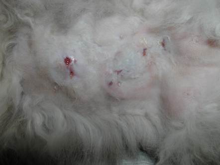
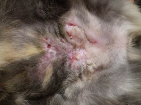
- Cutaneous lesions are characterized by dermal and subcutaneous nodules that evolve to develop ulcers and draining fistulae.
- Cats and Dogs:
-
-
- In the cat, lesions are more often seen on the caudal abdomen and inguinal region but other body regions, such as axillae and tail, can be affected.
-
-
-
- In the dog, lesions are more often present on the head.
- The lesions may or may not be painful and regional lymph nodes are not always enlarged.
- Fever and anorexia or other systemic signs are uncommonly observed with rapid-growing mycobacteria infections but can be seem in severely affected animals. However, infections with slow-growing organisms can more often disseminate and affect internal organs resulting in systemic signs.
- Cattle:
- Mycobacterium kansasii has been identified in cutaneous nodular lesions of cattle but in most cases the species involved are not determined.
- Skin lesions are characterized by papules and nodules, which often develop on the distal limbs and spread frequently to the thighs, shoulders and abdomen.
- Lesions can be single or multiple and frequently present in chains following the lymphatics.
- Lesions may drain a thick, cream to yellow to grayish pus.
-
-
Diagnosis
- History and clinical signs.
- Impression smears of exudate are often negative. Smears made from fine needle aspirates of closed lesions are more likely to contain the acid-fast organisms. Stain samples with acid-fast stains to increase the sensitivity to find the organisms. A Romanowsky stain is preferred by some and may show the negatively-stained organisms within macrophages.
- Bacterial culture and sensitivity are recommended because drug sensitivities can vary between mycobacterial species and even between isolates of the same species. Inform the laboratory that you are suspecting of mycobacteriosis because special culture medium such as Lowesnstein-Jensen or Stonebrink may be required.
- Biopsy samples should be stained with an acid-fast stain such as Ziehl-Neelsen or modified Fite-Faraco. It is recommended to cut a sample in three to four pieces or to take three to four small samples. One should be fixed in formalin for histopathology. One or two samples should be placed in a sterile container and frozen. If the specimen is acid-fast positive, one of the frozen pieces should be sent to a specialized laboratory for culture and sensitivity. One specimen should be kept frozen in case further investigation is needed.
- PCR should be considered in cases where culture and histopathology are unable to identify the microorganism. It can be done in fresh tissue or formalin-fixed, paraffin-embedded tissue. However, PCR is more sensitive if performed in fresh tissue.
-
Treatment
- Dogs and Cats:
- Wide surgical excision of the diseased tissue should be considered in all cases and may be curative.
- Long-term antibiotics should also be used.
- Ideally, antibiotics should be chosen based on culture and sensitivity results (very few laboratories in the U.S. will perform sensitivity for Mycobacterium spp.).
- When sensitivity data are not available, response may be seen to kanamycin, gentamicin, amikacin, chloramphenicol, tetracycline, doxycycline, potentiated sulfas, fluoroquinolones such as enrofloxacin, marbofloxacin and pradofloxacin, and clarithromycin. Combination antibiotic therapy is recommended to avoid resistance and increase the chances of success.
- Antibiotics should be used for 4 to 6 weeks after clinical cure. Extensive or systemic infections may require 6-12 months of therapy.
- The prognosis of opportunistic mycobacterial infections is always guarded, especially in cats.
- Cattle:
- The lesions of opportunistic mycobacteria infection cause little inconvenience, but they are unsightly.
- Treatment and control measures are typically not instituted, although surgical removal may be undertaken for cosmetic reasons.
- Dogs and Cats:
Important Facts
- Mycobacteria organisms are free-living, isolated from soil, decaying vegetation and water.
- Non-tuberculous opportunistic mycobacteria organisms are classified as rapid-growing and slow-growing.
- Trauma is necessary for infection to occur.
- Infection caused by rapid-growing mycobacteria is more often seen in cats.
- Dermal and subcutaneous nodules, which often ulcerate and fistulate are the classical lesions.
- Cats infected with rapid-growing organisms typically have lesions in the inguinal and abdomen regions because these sites have a thick layer of subcutaneous fat, which the mycobacteria organisms have tropism.
- In cattle, lesions are usually present on the limbs and in dogs on the head.
- Fever and anorexia or other systemic signs are uncommonly observed with rapid-growing mycobacteria infections.
- Infections caused by slow-growing opportunistic organisms can disseminate causing systemic signs.
- Diagnosis is made by finding acid-fast organisms in smears, cultures, biopsy specimens or molecular tests (PCR). Cytology and histopathology samples have to be stained with acid-fast stains to increase the sensitivity to find the organisms, which are scarce in rapid-growing mycobacteria infections.
- Treatment for dogs and cats involves surgical excision of affected tissue and long-term antibiotic therapy.
- Ideally, antibiotic should be selected based on culture and sensitivity.
- Combination antibiotic therapy is recommended to reduce the risk for resistance and increase the chance of success.
- Treatment is usually not instituted in cattle.
- Prognosis is guarded for cats and dogs, but is good for cattle.
References
Faccin M, Wiener DJ, Rech RR, et al. Common superficial and deep cutaneous bacterial infections in domestic animals: A review. Vet Pathol 2023; DOI: 10.1177/03009858231176558.
Gunn-Moore DA. Feline mycobacterial infections. Vet J 2014; 201: 230-238.
Malik R, Smits B, Reppas G, et al. Ulcerated and nonulcerated nontuberculous cutaneous mycobacterial granulomas in cats and dogs. Vet Dermatol 2013; 24: 146-e33.
Malik R, Wigney DI, Dawson D, et al. Infection of the subcutis and skin of cats with rapidly growing mycobacteria: a review of microbiological and clinical findings. J Fel Med Surg 2000; 2:35-48.
Scott DW. Bacterial Diseases. In: Large Animal Dermatology. Philadelphia, PA: W.B. Saunders, 1988; 146-150.
