3.18 Myiasis – Small Animals
Learning Objectives
- Know the types of myiasis that can affect small animals.
- Learn the predisposing factors associated with myiasis.
- Learn how to manage myiasis.
-
General Considerations
- Myiasis is the infestation of organs and tissues by larvae (maggots) of dipterous flies (order Diptera) that feed on necrotic or living tissue of the host.
- Myiasis is classified as facultative or obligatory.
- Most cases of myiasis in the U.S. are associated with larvae of flies that produce facultative myiasis.
- For facultative myiasis to develop there must be a factor such as traumatized skin, ocular discharge, discharge from any natural body orifice, neglected wound(s) or fecal/urine soiling to attract the female fly to deposit her eggs.
- Larval stages of facultative myiasis move over the wound surface ingesting secretions, exudate, dead cells, and debris but not live tissue as larvae of obligatory myiasis ingest. However, they induce irritation, injure cells, and provoke exudation.
- The fly of obligatory myiasis is dependent on fresh wounds as the site for larval development. These larvae can liquefy and devour viable tissues, thereby enlarging the wound.
- Obligatory myiasis occurs infrequently in the U.S. but are important in Central and South America and parts of Asia.
-
Cause
- Facultative Myiasis:
- Musca spp., Calliphora spp., Phaenicia spp., Lucilia spp., Phormia spp., and Sarcophaga spp.
- Obligatory Myiasis:
- Cochliomyia hominivorax (screwworm fly in the New World)
- Chrysomya bezziana (screwworm fly in the Old World)
- Facultative Myiasis:
-
Clinical Signs
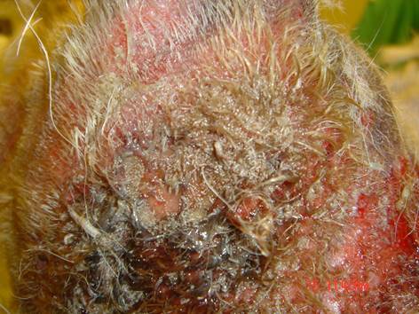
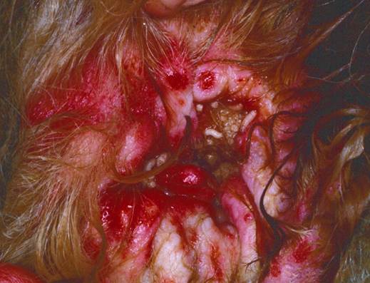
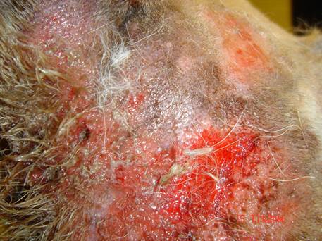
- Presence of larvae under matted hair and/or in wounds.
-
- They produce punched out lesions in the tissue and may tunnel in diseased tissues producing cavities.
-
- Often a foul odor is associated with the condition.
-
Treatment
- Various parasiticides have shown to be effective in the treatment of obligatory myiasis. Some examples include isoxazolines (fluralaner, afoxolaner, sarolaner, lotilaner), milbemycin, nitenpyram and spinosad. Combining some of these parasiticides may be more effective such as milbemycin and nitenpyram and milbemycin and spinosad.
- Hair and mats should be clipped from affected areas.
- Mechanical removal of the maggots using forceps and lesion cleansing with a diluted chlorhexidine solution are important part of the management.
- Necrotic tissue, if present, should be removed and the wound dressed with silver sulfadiazine to control infection.
- Owner should keep wound clean and, if possible, the animal should be housed in a fly free environment until healing occurs.
- Any secondary infection should be treated in an appropriate manner.
- Predisposing factors should be identified and controlled to prevent reoccurrence.
Important Facts
- Most cases of myiasis in the U.S. are caused by larvae of facultative flies. These larvae do not digest live tissue or enlarge the wound but cause wound irritation and exudation.
- Predisposing factors for myiasis to occur are traumatized skin, discharge from body orifices or from the eyes, and fecal/urine soiling.
- Clip the matted hair, clean the affected area, and remove all larvae with a forceps.
- Larvae causing obligatory myiasis typically need treatment with parasiticides such as isoxazolines, milbemycin, nitenpyram and spinosad.
- Identify and control the predisposing factor(s) to prevent reoccurrence.
References
Andriotti PA, Souza CP, Oliveira PC et al. Effectiveness of sarolaner in the clinical management of furuncular myiasis in dogs naturally infested with Dermatobia hominis (Diptera: Cuterebridae). Parasit Vectors 2021; 14:401.
Correia,TR, Scott FB, Verocai GG, et al. Larvicidal efficacy of nitenpyram on the treatment of myiasis caused by Cochliomyia hominivorax (Diptera: Calliphoridae) in dogs. Vet. Parasitol 2010; 173(1): 169-172.
Costa AJ, Martins JRS, Borges FA, et al. First report of the efficacy of a fluralaner-based pour-on product (Exzolt® 5%) against ectoparasites infesting cattle in Brazil. Parasit Vectors 2023; 16: 336.
Efficacy of afoxolaner (NexGard®) on the treatment of myiasis caused by the New World screwworm fly Cochliomyia hominivorax (Diptera: Calliphoridae) in naturally infested dogs. Vet Parasitol Reg Stud Reports 2021; doi: 10.1016/j.vprsr.2021.100569.
Siew Han H, Chen C and Schievano C, The comparative efficacy of afoxolaner, spinosad, milbemycin, spinosad plus milbemycin, and nitenpyram for the treatment of canine cutaneous myiasis. Vet Dermatol 2018; DOI: 10.1111/vde.12548.
Siew Han H, and Yasmin L. Chrysomya bezziana (Diptera: Calliphoridae) infestation in two Malaysian cats treated with oral lotilaner. Vet Dermatol 2020; DOI: 10.1111/vde.12855.
Scott DW, Miller WH, Griffin CE: Parasitic Skin Diseases. Small Animal Dermatology. 5th edn. Philadelphia: W.B. Saunders Co., 1995; 459-461.
Vale TL, Costa AR, Miranda LM et al. Efficacy of lotilaner against myiasis caused by Cochliomyia hominivorax (Diptera: Calliphoridae) in naturally infested dogs. Parasit Vectors 2023; 6: 86.
