2.8 Insect Bite Hypersensitivity – Horses
Learning Objectives
- Define the pathogenesis of insect bite hypersensitivity (IBH) in horses.
- What is the fly involved in equine IBH.
- Describe the classical history and clinical signs associated with IBH in horses.
- List the limitations of the skin biopsy and intradermal and serum allergen-specific IgE testing as diagnostic tools for IBH.
- Define the measurements involved in controlling flies.
- How would you use and when would you use antihistamines and glucocorticoids to manage IBH?
- What has been done to improve the efficacy of allergen-specific immunotherapy in horses with IBH?
- What are the future treatments for equine IBH?
-
General Considerations
- Insect bite hypersensitivity (IBH) is the most common allergic and pruritic skin disorder of horses. It is worldwide in distribution.
- It is seasonal (spring through fall) in temperate climates coinciding with the insect season. However, in tropical and subtropical climates it can be nonseasonal with exacerbation of signs during the insect season.
- It is a highly pruritic dermatosis and the hypersensitivity reaction is believed to be caused by the salivary antigens of the biting Culicoides spp. insects. The species of Culicoides involved vary around the world according to the geographic region.
- Culicoides midges prefer to feed during dawn and dusk, at which time hundreds of flies may bite a single horse.
- There is evidence that a hereditary predisposition to develop the hypersensitivity exists.
- The condition seems to worsen with age.
- A study noted that horses suffering from IBH have a higher risk for airway hyperreactivity and, therefore, might be predisposed to develop asthma in the future.
- Some horses with IBH may have concurrent atopic dermatitis and/or food allergy.
- Various other flies are known to bite horses including Simulium (black fly), Tabanidae (horse flies), Stomoxidae (stable flies), Culicidae (mosquitoes), and Phlebotominae (sandflies). However, these flies typically cause an inflammatory reaction at the biting site and not a hypersensitivity reaction.
- In the horse, the greatest loss caused by IBH is an aesthetic rather than economic value. There are monetary losses, however, to show or performance animals that cannot function as a result of their dermatitis.
Important Facts
- IBH is the most common cause of pruritus in horses.
- It is seasonal in temperate climates or year-round in tropical and subtropical climates.
- The disease is recurrent and becomes progressively worse with age.
-
Cause
- IBH is caused by bites of Culicoides spp midges, which are associated with the release of various salivary antigenic proteins. Studies have identified major salivary allergens to which at least 70% horses react. These studies have implications in the development of more accurate diagnostic tests and ultimately effective allergen-specific immunotherapy for IBH.
-
Pathogenesis
- A type I (IgE-mediated) hypersensitivity to various antigens present in the saliva of insects of Culicoides spp. midges is considered to play a central role in disease development. However, the pathomechanism is likely more complex and other parts of the immune system likely play a role. In fact, studies have shown a predominance of Th2 lymphocytes in the skin of IBH-affected horses. A Th1 response is considered protective against equine IBH.
- In horses that do not develop IBH, T-regulatory (FoxP3) lymphocytes appear to be involved in inducing tolerance to Culicoides saliva antigens. In contrast, horses with IBH have a decrease in T-regulatory lymphocytes.
- IL-31 appears to play a major role in the pruritus associated with IBH, evidenced by the increased expression of IL-31 mRNA and its receptor in the skin of affected horses. These findings have important implications in the treatment of this disease.
- A study showed that the skin of affected horses has an altered epithelial barrier which may act as a predisposing factor for the development of IBH.
Important Facts
- Type I (IgE mediated) hypersensitivity to various antigens in the saliva of Culicoides spp. midges.
- IL-31 appears to play a major role in the pruritus associated with IBH.
-
Clinical Signs
- Since the disease is allergic in nature, its occurrence is sporadic, i.e. usually only one animal out of a stable or pasture will be affected.
- IBH is rare or nonexistent in horses under one year of age. Most horses will develop clinical signs at the age of 3 to 4 years. Horses exposed to bites of Culicoides spp. midges later in life are more prone to develop IBH.
- There is no age or sex predilection; however, IBH appears to be more frequent in some breeds such as, Icelandic, German Shire, Belgian warmblood, Dutch Shetland pony, Arabians, Connemaras, and quarter horses. It is widely accepted that horses are genetically predisposed to develop IBH and that it is a polygenetic disease.
- In northern climates where there is a marked difference in ambient temperature, the “allergy season” is distinct. The condition is recurrent and with each succeeding year the signs become more severe and the season gets longer. The seasonality directly corresponds to the activity levels of the Culicoides spp. midges.
- In southern climates where there is not a distinct cold winter, theCulicoides spp. midges and, therefore, the hypersensitivity disorder are present year round but the condition will be worse when the insect population is greater.
- Clinical signs may be worse near dusk and dawn, which are the favorite feeding times for Culicoides spp. midges.
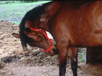
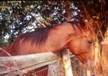
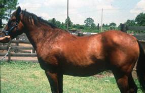
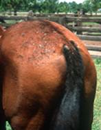
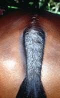
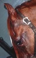
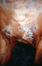
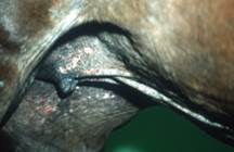
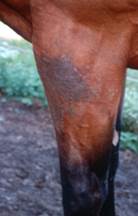
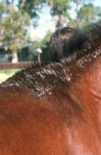
- Pruritus, crusted papules and urticaria are the initial clinical signs. As the disease becomes chronic, excoriations, self-induced alopecia, lichenification, pigment disturbances, a “rat tail” and a “buzzed off” mane will develop.
-
- The distribution of lesions typically correlates with the feeding sites of the Culicoides midges present in the geographical region. It can vary as follows:
- Dorsal distribution: usually begins at the mane, rump, and base of the tail. Then, it progresses to affect the face, pinna, neck, shoulder and dorsal thorax.
- The distribution of lesions typically correlates with the feeding sites of the Culicoides midges present in the geographical region. It can vary as follows:
-
-
- Ventral distribution: ventral thorax and ventral abdomen, axilla, and groin. Legs and intermandibular area are often affected.
-
-
-
- Combination: features of dorsal and ventral surface.
- In some cases, the lesions are limited to the mane and withers regions.
-
-
-
- Flanks are rarely affected.
- Kicking at flies or running off can lead to injury to the horse and owner.
-
Important Facts
- IBH is rare in horses less than one year of age.
- It typically affects one animal out of a stable or pasture supporting its allergic nature.
- Pruritus and crusted papules are the initial signs. As the disease progresses excoriations, alopecia, lichenification, pigment disturbances, a “rat tail” and a “buzzed off” mane are seen.
- Base of the tail, rump, back, withers, crest, poll and ears are classically affected. However, ventral distribution with or without involvement of the mane and tail can also be seen.
- Lesions can be generalized.
-
Diagnosis
- The history and clinical signs are very suggestive.
- Rule out other pruritic skin disorders that typically affect one horse in the farm such as atopic dermatitis and food allergy. If multiple horses are pruritic consider mange, pediculosis, and insect bite dermatitis.
- For pruritus localized to the tail, consider pinworm infestation (oxyuriasis), pediculosis (lice), chorioptic mange, atopic dermatitis, food allergy, stable vice and tail pyoderma. Keep in mind that for most of these disorders, with the exception of stable vice and oxyuriasis, typically pruritus will also be present in other parts of the body.
- Skin scrapings are helpful in ruling out ectoparasite problems (e.g., e.g. chorioptic and psoroptic mange).
- Skin cytology and culture are useful in determining whether a bacterial (e.g. Staphylococcus sp., Dermatophilus congolensis) or fungal (Trichophyton mentagrophytes, Microsporum canis, Microsporum gypseum) infection is present.
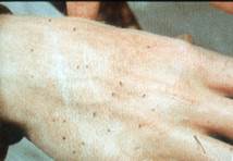
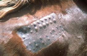
- Intradermal (IDT) or serum allergen-specific IgE testing can be used to support a clinical diagnosis of IBH. Ideally, IDT reactions should be read at 30 minutes, 4 hours and 24 to 48 hours after the test. However, the sensitivity of the test may not increase significantly with readings after 30 minutes. It is important to keep in mind that normal horses may have positive reactions to Culicoides spp antigens especially at 30 minutes after testing. It is also important to keep in mind that false positive and false negative reactions also occur with the serum allergen-specific IgE test. Advances in molecular testing using recombinants of Culicoides major allergens are already improving the sensitivity and specificity of these tests.
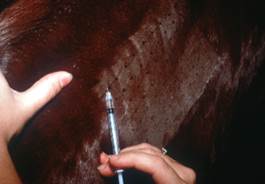
Testing the lateral aspect of the neck of a horse with suspected Culicoides spp. hypersensitivity
-
- Skin biopsies results can support a presumptive diagnosis but findings are non-specific. Histopathology most often demonstrates superficial and deep perivascular dermatitis with eosinophils and mononuclear cells. This inflammatory pattern is suggestive of IBH but not diagnostic because it can also be seen in atopic dermatitis and food allergy.
- A favorable response to adequate insect control measures supports a clinical diagnosis.
Important Facts
- Diagnosis of IBH is usually based on a characteristic history and clinical signs.
- Molecular tests using recombinant proteins of Culicoides major allergens have improved the sensitivity and specificity of the allergen-specific intradermal and serologic tests in the diagnosis of IBH.
-
Treatment
- The primary objective in treating IBH dermatitis is avoidance or reduction of exposure to Culicoides spp. midges.
- Reduction of exposure can be attempted in a few ways:
- Application of residual insecticides and repellents (must be used frequently and religiously) such as, pyrethroids (permethrin appears to be the best choice); Avon-Skin-So-Soft (repellent); Ceratex® (DEET-repellent). It can be applied on the animal in addition to the housing and screens.
- Stabling during peak feeding times of Culicoides spp. (i.e. from dusk until dawn).
- Culicoides spp. midges are very small; therefore, a smaller mesh screen than the one used for mosquitoes have to be applied.
- Powerful fans (overhead or floor) can be used in stables because Culicoides spp. are weak fliers.
- Elimination of larval development areas: compost, standing water, understory of brush.
- When avoidance is not practical or the offending agent is not known, antihistamines and/or systemic glucocorticoid therapy can be instituted.
- Antihistamine: hydroxyzine at 1 to 2 mg/kg q 8-12h orally; chlorpheniramine at 0.25 to 0.5 mg/kg q 12h orally; diphenhydramine at 1 to 2 mg/kg q 8-12h orally; and doxepin at 0.5 to 0.75 mg/kg q 12h orally.
- Glucocorticoid: Prednisolone at the dose of 1.0 to 2.2 mg/kg orally and daily should be given until pruritus and secondary self-trauma are controlled, then the dosage should be tapered to the lowest dose that will control the disease when given every other day. Dexamethasone at 0.2 mg/kg q 24h can also be used.
- Topical antipruritic cream or lotion such as products containing 1% hydrocortisone acetate and 1% pramoxine HCL can be tried.
- Immunotherapy based on intradermal or serum allergen-specific IgE testing should be considered as part of the treatment regimen. Researchers have identified some of the major Culicoides spp allergens and allergen-specific immunotherapy using these allergens have been effective in controlling the hypersensitivity in affected horses. As more research are done using major allergens in the immunotherapy, it could become the mainstay treatment for IBH.
- If the horse does not show any response after 12 months of treatment, immunotherapy will probably not work.
- A vaccine against equine IL-5 bound to virus-like particle (VLP) was developed with the goal of inducing neutralizing IL-5 antibodies which is a cytokine involved in the migration, activation and survival of eosinophils. Preliminary results showed that it is safe and effective with 47% of the treated horses having 50% improvement in clinical scores and 21% having 75% improvement.
- Using a similar approach, a vaccine using IL-31 was also developed to induce neutralizing antibodies against IL-31 and, therefore, reduce the pruritus associated with IBH. A double-blind, placebo-controlled, randomized trial showed a reduction in lesion scores in IBH horses compared to placebo.
Important Facts
- The primary objective in treating IBH dermatitis is avoidance or reduction of exposure to Culicoides spp midges.
- When avoidance is not practical, antihistamines and/or systemic glucocorticoid therapy can be instituted.
- Allergen-specific immunotherapy using recombinant proteins of major Culicoides spp allergens that reacted on the intradermal or serum allergen-specific IgE test, should be considered as part of the treatment regimen. As more studies are conducted investigating the efficacy of allergen-specific immunotherapy using major salivary allergens, this treatment modality may become the mainstay of IBH therapy.
- Vaccines using IL-5 and IL-31 have been developed and preliminary studies have indicated that they are safe and have improved the IBH clinical signs.
References
Cox A, Stewart AJ. Insect bite hypersensitivity in horses: Causes, diagnosis, scoring and new therapies. Animals 2023; doi.org/10.3390/ani13152514.
Cvitas I., Oberhansli S, Leeb T et al. Investigating the epithelial barrier and immune signatures in the pathogenesis of equine insect bite hypersensitivity. PLOS ONE 2020; 15(4): e0232189.
Fadok VA. Update on equine allergies. Vet Clin Equine 2013; 29: 541–550.
Fettelschoss-Gabriel A, Birkmann K, Pantelyushin S, et al. Molecular mechanisms and treatment modalities in equine Culicoides hypersensitivity. Vet J 2021; 276: 105741.
Fettelschoss-Gabriel A., Fettelschoss V, Thoms F. et al. Treating insect-bite hypersensitivity in horses with active vaccination against Il-5. J Allergy clin Immunol 2018; 142 (4): 1194-1204.
Lanz S, Brunner A, Graubner C et al. Insect Bite Hypersensitivity in Horses is Associated with Airway Hyperreactivity. J Vet Intern Med 2017:1-7.
Marsella R. Pruritic horse: Approach to allergic skin diseases in horses. Vet Clin Equine 2024; 40:219-235.
Marcella R, White S, Fadok VA, et al. Equine allergic skin diseases: Clinical consensus guidelines of the World Association for Veterinary Dermatology. Vet Derm 2023; 34: 175-208.
Marti E, Novotny EN, Cvitas I et al. Immunopathogenesis and immunotherapy of Culicoides hypersensitivity in horses: an update. Vet Derm 2021; 32: 579–e156.
Scott, DW. Large Animal Dermatology. In: Immunologic Diseases. W.B. Saunders, Philadelphia, PA, 1988, p 300 – 301.
Schaffartzik A., Hamza E. Janda J. et al. Equine insect bite hypersensitivity: What do we know? Vet Immunol Immunopathol 2012; 147: 113-126.
Yu AA. Equine Allergies: Insect Hypersensitivity. In Proceedings of the Ontario VMA Conference & Trade Show : Ontario, VMA, USA: 2016.
