1.9 Exudative Epidermitis – Large Animals
Learning Objectives
- What organism causes most cases of exudative epidermitis or greasy pig disease?
- At what age are the animals affected?
- Describe the three clinical forms of the disease.
- How do you diagnose exudative epidermitis?
- How do you treat this condition?
-
General Considerations
- Exudative epidermitis or greasy pig disease is typically a disease of suckling or newly weaned pigs.
- Affected piglets may die of anorexia and dehydration a few days after the infection.
- More than one piglet is typically affected in a farm and the disease can be a significant cause of mortality and poor growth rate.
-
- The disease has a worldwide distribution.
-
Etiology
- Staphylococcus hyicus subsp. hyicus is by far the most common agent; however, other staphylococci capable of producing exfoliative toxins including S. sciuri, S. chromogenes and S. aureus have been rarely isolated from skin lesions.
-
Clinical Signs
- Clinically, the disease is divided into peracute, acute and subacute forms.
- Peracute:
- Initially, a dark-brown “waxy” crust develops periocularly, and then a vesicopustular eruption develops on the nose, lips, tongue, gums, and coronets.
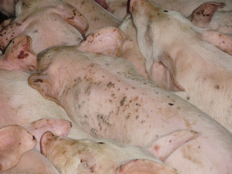
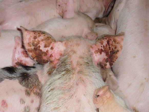
- The disease progresses to affect the entire body with lesions characterized by erythema, a moist to greasy exudate and brown crusts.
- The feet often presents erosions of the coronary band and heel.
- The piglets show progressive depression, anorexia, and polydipsia and usually die within 3 to 5 days.
- Internal organs may also be affected.
- Peracute:
- Clinically, the disease is divided into peracute, acute and subacute forms.
-
- Acute:
- The clinical presentation is similar to the peracute form but the skin becomes thicker and wrinkled in the acute form.
- Death typically occurs within 4 to 8 days.
- Subacute:
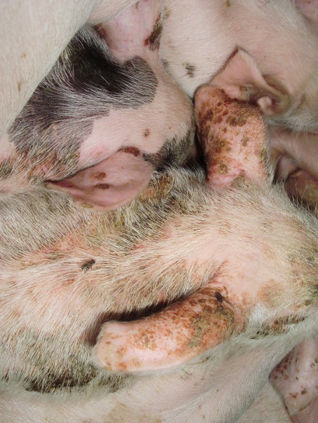
- Skin lesions are milder and typically limited to the head and ears and are less exudative.
- The piglets are generally healthy otherwise and the disease resolves spontaneously.
- Acute:
-
Diagnosis
- History and characteristic clinical signs.
- Differential diagnoses include sarcoptic mange, dermatophytosis, pityriasis rosea, zinc deficiency, swine pox and biotin deficiency.
- Cytology of exudative lesions shows coccoid-shaped bacteria.
- Biopsy should be performed to confirm a presumptive clinical diagnosis and to rule other diseases. It shows subcorneal vesicular-to-pustular dermatitis and acantholytic keratinocytes.
- Culture from vesicles or pustules and conjunctiva yields S. hyicus and, less frequently, the other staphylococci species mentioned above.
-
Treatment and Prognosis
- The worse the clinical condition the smaller the chances of response to treatment.
- Ideally, antibiotic therapy should be based on culture and sensitivity results as resistant strains of S. hyicus have been reported.
- Penicillin (5000 IU/Kg twice daily) given IM for 3 to 5 days has been effective.
- Tylosin (8 mg/kg) given IM for 2 to 3 days has also been reported to be beneficial.
- Good results have been seen with one injection of oxytetracycline (11 to 18 mg/kg) followed by oxytetracycline in the feed (300g/Ton).
- Good hygiene and management practices should be instituted to prevent infection.
- Immunization has been reported to be effective in the prevention of experimentally induced exudative epidermitis.
- The peracute and acute forms of the disease are severe and often result in death. In contrast, the subacute form typically spontaneously resolves.
Important Facts
- Exudative epidermitis or greasy pig disease is a disease of suckling or newly weaned pigs.
- It is typically caused by Staphylococcus hyicus subsp. hyicus.
- Adhered dark-brown crusts and greasy exudate are characteristic clinical signs.
- The diagnosis is based on history, clinical signs, biopsy and bacterial culture.
- The response to treatment correlates inversely with the duration and form of the infection.
- The mainstay treatment is antibiotic therapy.
- Ideally, the antibiotic should be selected based on culture and sensitivity results.
- The peracute and acute forms of the disease often result in death.
References
Foster AP. Staphyloccal skin disease in livestock. Vet Dermatol 2012; 23:342-352.
Lua L, Hea K, Nia Y et al. Exudative epidermitis of piglets caused by non-toxigenic Staphylococcus sciuri. Vet Microbiol 2017; 199:79-84.
Mebus CA, Underdahl NR, Twiehaus, MJ. Exudative epidermitis: pathogenesis and pathology. Vet Pathol 1968; 5: 146-163.
Scott DW. Bacterial Diseases. In: Large Animal Dermatology. Philadelphia, PA: W.B. Saunders, 1988; 123-126.
Wegener HC, Skov-Jensen EW. Exudative epidermatitis. In: Straw BE, Zimmerman JJ, D’Allaire S et al., eds. Diseases of Swine, 9th edn. Ames, IA: Blackwell Publishing, 2006; 675-679.
