4.2 Dermatophytosis – Large Animals
Learning Objectives
- Know that horses under 4 years of age and cattle under 1 year of age are predisposed to develop dermatophytosis.
- Remember! Dermatophytosis is a zoonosis.
- Learn the clinical signs of dermatophytosis in horses and cattle.
- Remember! Fungal culture is the most accurate diagnostic test. However, go ahead and perform also a direct examination of hairs and scales. Do not rule out dermatophytosis if the direct examination test is negative.
- Describe the technique to collect samples for fungal culture and direct examination.
- Describe the technique of the direct examination test.
- Remember! Trichophyton equinum and Trichophyton verrucosum require vitamin enrichment medium to grow.
- Remember! Dermatophytosis is a self-limiting condition. Know the reasons for treating dermatophytosis.
- Learn the treatment options for large animals.
- Learn the cases that can benefit from vaccination.
- Learn the recommendations to prevent infection and/or re-infection.
-
General Considerations
- Dermatophytosis is a fungal infection of the keratinized skin structures including the stratum corneum, hairs, and hooves.
- It occurs more often in horses, cattle, goats and swine but it is uncommon in sheep.
- The most important mode of transmission is the direct contact with affected animals and less commonly by contact with infected hairs and crusts on fomites or in the environment.
- Although of limited morbidity, dermatophytosis can be problematic because of the potential for outbreaks, prolonged disease (at the individual or herd levels), cost, bother of treatment, and the potential for human infection.
- Horses under 4 years of age, cattle less than 1 year old and lambs are more susceptible due to lack of immunity.
- It occurs most frequently in the winter months and especially in confined animals.
- Dermatophytosis is a zoonosis.
Important Facts
- Dermatophytosis is common in horses, cattle, goats and swine but it is uncommon in sheep.
- It occurs more often in horses under 4 years of age, cattle less than one year old and lambs.
- It is a zoonosis.
-
Etiology and Pathogenesis
- Species important in the horse include: Trichophyton equinum (most common); Trichophyton mentagrophytes; Trichophyton verrucosum, Microsporum equinum, Microsporum gypseum; and Microsporum canis. In horses, Trichophyton spp have shown to produce proteolytic enzymes that induce keratinocyte acantholysis in vitro and in vivo (lesions can suggest pemphigus foliaceus on cytology and skin biopsy).
- Most common species in cattle and goats: Trichophyton verrucosum; Trichophyton mentagrophytes.
- Species important in sheep include: Trichophyton mentagrophytes, Trychophyton verrucosum, Microsporum gypseum, and Microsporum canis.
- In swine, Microsporum nanum is the most common cause of dermatophytosis with Trichophyton mentagrophytes, Trichophyton verrucosum and Microsporum canis being less commonly isolated.
-
Clinical Signs
- Equine:
- The clinical features of typical dermatophytosis in the horse include:
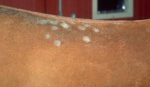
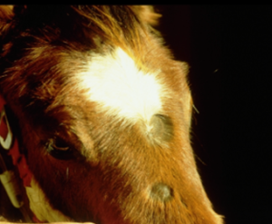
- Focal to multifocal areas of round to oval well-demarcated alopecia, scaling and/or crusting.
- The clinical features of typical dermatophytosis in the horse include:
- Equine:
-
-
-
- Pruritus is uncommon but may be present in some animals.
-
-
-
-
- The lesions are almost always multiple and occur most commonly in the saddle and girth region.
- Initially, the lesions may appear as urticarial-like swellings. Soon after that, the hair comes out easily in the affected areas and the alopecic areas develop scales and/or crusts.
- Annular and coalescing lesions expanding centrifugally and losing their circular appearance are signs reported in horses with Trichophyton spp. infection.
-
-
-
- “Atypical” forms of equine dermatophytosis may occur.
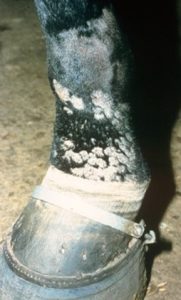
- Infection with Microsporum gypseum tends to be associated with much thicker crusts, more inflammation, suppuration and ulceration.
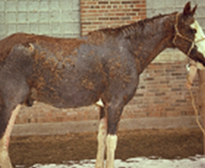
- In some horses, the disease presents as extensive scaling and crusting without significant alopecia.
- Occasionally, the lesions are limited to the pastern region and are erroneously diagnosed as “scratches” or “grease heel”.
- “Atypical” forms of equine dermatophytosis may occur.
-
-
-
- Consider dermatophytosis as a possible differential diagnosis in any horse with skin disease because of its variable clinical presentation and potential for rapid spread.
-
Important Facts
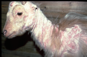
- Typical lesions of dermatophytosis in horses are characterized by multifocal areas of alopecia, scaling and crusting.
- In horses, lesions are often seen in the saddle and girth regions.
- Initially, lesions may look like urticarial-like swellings.
-
- Bovine:
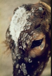
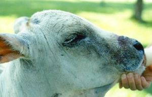
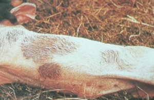
- Trichophyton verrucosum – lesions usually develop around the head and neck and are typically associated with a thick keratotic crust.
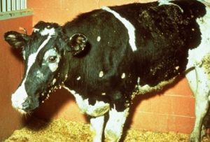
- Trichophyton mentagrophytes – lesions are characterized by multiple 1/2 to 1 cm areas of focal alopecia with minimal scaling, they are not crusty.
- Bovine:
-
- Sheep:
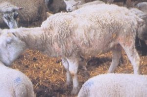
- Lesions are commonly seen on the face, neck, thorax and back and are characterized by circular areas of alopecia and thick grayish crusts.
- Sheep:
-
- Goat:
- Lesions are most commonly seen on the face, pinnae, neck and limbs.
- They are typically circular to diffuse areas of alopecia, scaling, erythema, and yellowish-gray crusts.
- Goat:
-
- Swine:
- Lesions are most commonly seen behind the ears and on the trunk and are characterized by annular areas of red to brown discoloration of the skin and superficial dry-brown to orange crusts.
- Swine:
-
-
- Alopecia and pruritus are rare.
-
-
Diagnosis
- The diagnosis of dermatophytosis is frequently given based on the clinical appearance of lesions; however, it should be confirmed with one or more of the diagnostic tests listed below.
- Wood’s lamp.
- Limited value as a diagnostic tool because of the relatively high number of false positive and negative results.
- Microsporum canis and Microsporum equinum are the only dermatophytes affecting food animals and horses that will produce fluorescence. In addition, not all strains of these dermatophyte species fluoresce.
- Trichogram or direct microscopic examination of hairs has low sensitivity. Therefore, do not rule out dermatophytosis if this test is negative. Pluck the abnormal hairs localized within the lesion and close to the periphery. However, if abnormal/broken hairs are only present at the center of the lesion, do not hesitate to pluck them.
- Biopsy is rarely needed but should be considered in cases with unusual clinical signs. Special stains may be necessary to visualize the fungal elements (PAS and/or GMS).
- Fungal Cultures.
- The most definitive method of diagnosis but results will be negative if samples are not collected properly.
- It identifies the source of infection.
- Use the technique described for small animals. Pluck the abnormal hairs localized within the lesions and close to the periphery. However, if abnormal/broken hairs are only present at the center of the lesion, do not hesitate to pluck them.
- Vitamin enrichment media is needed to culture Trichophyton equinum and Trichophyton verrucosum. Trichophyton equinum requires nicotinic acid and Trichophyton verrucosum requires thiamine and inositol.
Important Facts
- Remember, only a proportion of Microsporum canis and Microsporum equinum strains fluoresce under the Wood’s lamp! Thus, if no fluorescence is noted, do not rule out a possible diagnosis of dermatophytosis.
- Fungal culture is a reliable diagnostic tool if samples are collected properly.
- Pluck the abnormal hairs localized within the lesion and close to the periphery. However, if abnormal/broken hairs are only present at the center of the lesion, do not hesitate to pluck them.
- Trichophyton equinum requires nicotinic acid and Trichophyton verrucosum requires thiamine and inositol to grow in culture media.
-
Treatment
- Most cases will resolve spontaneously in 1 to 3 months unless the animal is debilitated by illness, parasite infection, malnutrition, etc.
- The goal of the treatment is to decrease the course and severity of the disease, prevent infection spread, and decrease environmental contamination.
- Good nutrition and adequate exposure to sunlight is advised.
- Treatment includes fungicidal therapy for the animal and decontamination of the environment and fomites (tack, brushes, blankets, etc.). Clorox® (bleach) can be used in the environment.
- All in-contact animals should be treated.
- Topical fungicides most often recommended include:
- Lime sulfur 3 to 5% solution.
- Enilconazole, a beta-substituted imidazole, shows excellent efficacy against dermatophytes. It is currently used in Europe as a topical and environmental fungicide for large and small animals. It is currently available in the US as an environmental fungicide for poultry industry (Clinafarm® EC).
- A study used 0.2% Clinafarm® EC topically (i.e. Clinafarm® EC – 14.5 ml of the 13.8% concentrate added to 1 L of water) as adjunctive therapy for feline dermatophytosis with good results. The solution was applied as a rinse.
- Povidone iodine; can irritate and dry the skin.
- Chlorhexidine 3% or 4 % solution.
- Benzalkonium chloride 0.6% solution.
- These products are used as a dip or rinse.
- Topical fungicides should be applied daily with a sponge or brush to the entire body for one week then twice weekly until clinical resolution.
- All crusts should be removed to facilitate treatment and disposed off carefully.
- In horses, creams or ointments containing miconazole, clotrimazole, terbinafine or luliconazole can be used in the affected areas.
- Treatment should be continued for at least 6-8 weeks. A good rule-of–thumb is to treat for 2 weeks past clinical resolution but ideally treatment should not be discontinued until at least one negative culture is obtained.
- Handle affected animals last.
- Systemic therapy with griseofulvin:
- Actual efficacy is unknown due to high rate of self-cure.
- Very costly.
- Teratogenic – do not use in pregnant animals.
- 60 mg/kg q 24h for 5 weeks was effective in calves.
- In the horse, 10 mg/kg q 24h for at least 30 days has been suggested.
- Considering the cost and potential toxicity of oral antifungal medications, combined with the self-limiting nature of disease and relative ease of topical therapy; topical therapy alone is a reasonable approach, with oral treatment reserved for refractory or perhaps severe cases. Moreover, the pharmacokinetic of systemic antifungals has not been studied in food animals and horses.
Important Facts
- Most cases will resolve spontaneously in 1 to 3 months unless the animal is debilitated by illness, parasite infection, and malnutrition.
- The treatment goal is to decrease disease course and severity, prevent the spread of infection, and reduce environmental contamination.
- Treatment includes fungicidal therapy (primarily topical) and decontamination of the environment (tack, brushes, blankets, etc).
- Treat all in-contact animals.
- Affected animals should be handled last.
-
- Vaccination – used in Europe.
- Trichophyton verrucosum modified live vaccine.
- Life-long immunity.
- Calves are vaccinated at 2 weeks of age.
- No apparent interference of maternal antibodies.
- Not protective against other dermatophytes.
- Experimental Trichophyton equinum vaccine in horses was 91% effective. Horses that developed ringworm only developed single lesions.
- Consider vaccination in chronic and recurrent epidemics.
- Trichophyton verrucosum modified live vaccine.
- Prevention:
- Do not share tack or brushes.
- Routine disinfection of equipment.
- Avoid overcrowding.
- Good health-care practices.
- General infection control practices including quarantine of new arrivals and prompt diagnosis of suspected cases are important.
- Prophylactic topical treatment of exposed animals should be considered.
- Vaccination – used in Europe.
References
2013; 167: 215-234.
Hnilica and Medleau. Evaluation of topically applied enilconazole for the treatment of dermatophytosis in a Persian cattery. Vet Dermatol 2002; 13:23-28.
Scott, DW. Large Animal Dermatology. In: Fungal Diseases. W.B. Saunders, Philadelphia, PA, 1988, p 172 – 182.
2021; 32: 474-e129.
Weese JS, Anthony A Y. Infectious folliculitis and dermatophytosis. Vet Clin North Am Equine Pract. 2013; 29: 559-575.
