2.7 Contact Dermatitis – Small and Large Animals
Learning Objectives
- Know the two forms of contact dermatitis (CD).
- Understand the pathogenesis of CD.
- Be aware of the potential allergens and irritants that can trigger CD.
- Describe the clinical signs of CD.
- Be familiar with the diagnostic tests of CD.
- Learn how to treat CD.
-
General Considerations
- Contact dermatitis is an inflammatory and pruritic skin response to the contact of an antigenic or irritant substance.
- It is an uncommon skin disease of domesticated animals but its occurrence should not be underestimated.
- There are two forms of contact dermatitis: allergic contact dermatitis (ACD) and irritant contact dermatitis (ICD). ACD is an allergen-specific immunologic response to an allergenic substance and is not dose dependent. In contrast, ICD is a nonspecific inflammatory response to an irritating substance and it is dose dependent.
- Moisture is an important predisposing factor because it decreases the effectiveness of the normal skin barrier and increases the intimacy of contact between the substance and the skin surface.
- It is possible that contact dermatitis is underdiagnosed because an accurate diagnosis is difficult to achieve.
Important Facts
- Contact dermatitis is uncommon in domestic animals but it is possible that it is underdiagnosed because an accurate diagnosis is difficult to achieve.
- Contact dermatitis can be allergic or irritant.
-
Pathogenesis
- ACD – the pathomechanism involves a type IV (delayed, cell mediated) hypersensitivity reaction that develops in previously sensitized individuals. Antigen-presenting cells and cytotoxic CD8+ T-lymphocytes are the central players.
- Two phases participate in the pathogenesis of ACD: the sensitization phase and elicitation phase. In the sensitization phase, the immune system becomes sensitized to a specific antigen (sensitizer). It is followed by the elicitation phase where the immune system triggers an antigen-specific response after re-exposure to the sensitizer.
- The sensitizer is usually a hapten, which becomes a complete antigen upon union with an epidermal protein. The sensitizer-protein complex is required for the substance to become an allergen.
- The sensitization phase can last weeks to months of intermittent or continual exposure to the sensitizer. During sensitization, epidermal Langerhans cells or dermal dendritic cells processes the sensitizer-protein complex and presents the processed allergen to allergen-specific lymphocytes. This will result in clonal expansion of these allergen-specific lymphocytes.
- After sensitization has occurred the clone of allergen-specific lymphocytes will respond within 24 hours to allergen re-exposure resulting in the release of various pro-inflammatory cytokines and chemokines. This characterizes the elicitation phase.
- An interesting thing to keep in mind is that most sensitizers are also irritants. In this case, an inflammatory response can develop before the sensitization phase is completed.
- Potential allergens for dogs and cats include:
- Plants (e.g. Commelinaceae family [e.g. wandering Jew plant, spreading dayflower], Asian jasmine, Amarylidaceae leaves and bulbs).
- Grass leaf.
- Topical medications (e.g. neomycin).
- Industrial materials (e.g. plastic, cement, wood).
- Vehicles for topicals (e.g. propylene glycol).
- Skin products (e.g. shampoos, dips, sprays).
- Potential allergens for large animals:
- The most commonly incriminated contact sensitizers in horses include pasture plants (e.g. buttercups), bedding, soaps, insect repellents, topical medications, and tack items (dyes and preservatives)
- ICD – After the skin barrier is disrupted, an irritant substance an irritant substance causes cytotoxic damage to keratinocytes when it contacts the skin resulting in the release of various inflammatory mediators. Irritants are directly responsible for the inflammatory reaction and lesions of ICD. Prior exposure to the irritant substance and sensitization are not required. The lesion severity depends, at least to some extent, on the substance concentration, potency and contact time.
- Potential irritants:
- Strong acids and alkali.
- Cleansers (e.g. soaps, detergents).
- Solvents.
- Insecticides (e.g. flea collars, flea shampoos, flea dips).
- Grasses and pollens.
- Industrial chemicals (e.g. fertilizers, petrolatum).
- Industrial materials (e.g. paint, carpets, wood preservatives).
Important Facts
- In allergic contact dermatitis (ACD) an allergen-specific immunologic response develops to an allergenic substance and the reaction is not dose dependent. Pre-exposure is needed for sensitization to occur.
- Irritant contact dermatitis (ICD) is a nonspecific inflammatory response to an irritating substance and it is dose dependent. Pre-exposure is not required for the reaction to occur.
- Potential allergens include medications, grass and plants, plastic, cement, wood, medicated shampoos, dips and sprays, insect repellents, bedding, and propylene glycol.
- Potential irritants include strong acids and alkali, soaps, detergents, solvents, insecticides, grasses, pollens, fertilizers, petrolatum, paint, carpets, and wood preservatives.
- An interesting thing to keep in mind is that most sensitizers are also irritants.
-
Clinical Signs
- Small Animals:
- Mild to moderate pruritus – not as intense as the lesions might suggest.
- Areas involved:
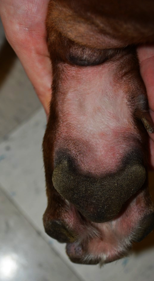
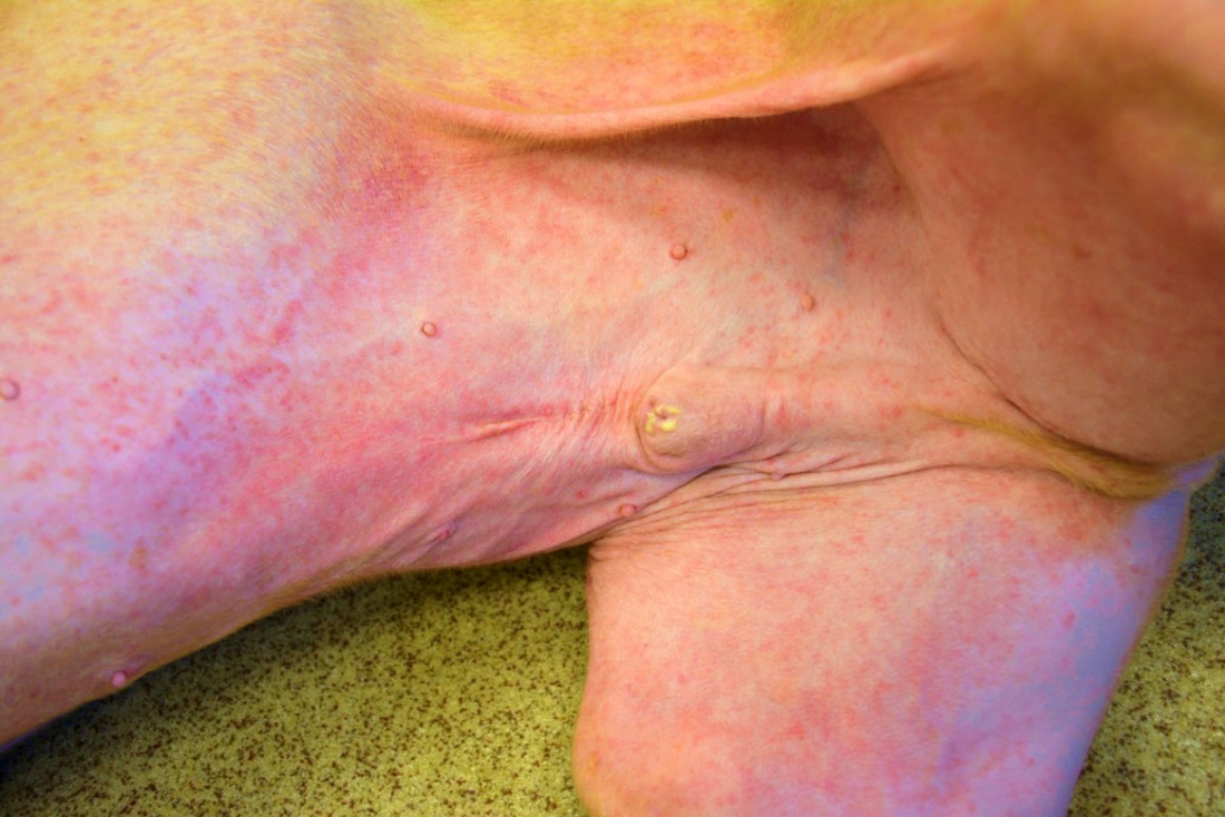
- Contact areas, which include the thinly haired or hairless skin such as ventral abdomen, scrotum, volar aspect of the paw, axilla, groin, perineum, and brisket area. This distribution can mimic food-induced or environmental-induced atopic dermatitis.
- Small Animals:
-
-
-
- If the haired parts of the trunk are diffusely affected, consider a reaction to shampoo. Lesions localized between the shoulder blades should increase the index of suspicion for a reaction to a spot-on anti-parasitic product.
- Lesions at the concave aspect of the pinna are usually from topical medications.
- Lesions caused by an irritant substance tent to be limited to the area of direct contact and are often well demarcated.
- Lesions caused by an allergen tent to extend beyond the area of contact.
- What are the lesions?
- Acute lesions include papular eruptions, erythema and edema.
-
-
-
-
-
- Chronic lesions include lichenification, hyperpigmentation, scaling and fissuring.
- ACD is more likely to induce vesicles and ICD is more likely to induce epidermal necrosis and ulceration
- Secondary superficial pyoderma and/or bacterial and yeast overgrowth/dermatitis may occur.
-
- Large Animals:
- Contact dermatitis has been reported more often in horses than other large animal species.
- Pruritus is variable.
- The area of the body affected suggests the contactant:
- Face, ears, and neck: insect repellents and sprays.
- Muzzle and extremities: pasture and bedding.
- Face and trunk: tack.
- Because of the horse’s ability to sweat profusely, contactants have access to the skin even in heavily haired areas.
- What are the lesions?
- Acute lesions include papules or vesicles, erythema, edema, exudation.
- Chronic lesions include self-induced alopecia, lichenification, hyperpigmentation.
- General facts:
- ICD typically affects multiple individuals in the household or farm. In contrast, ACD typically affects a single individual.
- Lesions of ICD and ACD overlap making it difficult to achieve a diagnosis based solely on lesion characteristics.
- Pain and discomfort is generally associated with ICD whereas pruritus is associated with ACD.
-
Important Facts
- Lesions typically develop in thinly or hairless areas; however, think of a shampoo reaction or a spot-on anti-parasitic reaction if lesions develop diffusely throughout the trunk or in between the shoulder blades, respectively.
- Acute lesions include papules, vesicles and edema.
- Chronic lesions include hyperpigmentation, lichenification, scaling and fissures.
- Vesicles are more likely to develop with ACD and epidermal necrosis and ulceration with ICD.
- Pain and discomfort is usually associated with ICD and pruritus with ACD.
- Lesions of ICD and ACD overlap making it difficult to achieve a diagnosis based solely on lesion characteristics.
-
Diagnosis
- The patient’s history and clinical signs (contact areas involved) are very important parts of the diagnostic equation.
- A detailed history is extremely important. Remember, most substances that cause contact dermatitis are not new in the environment. Some questions that may help differentiate between ACD and ICD include previous CD events, number of animals affected, medications used, contact to grass, plants, cement, plastic, insecticides used etc.
- Major differential diagnoses include food-induced or environmental-induced atopic dermatitis, adverse drug reactions, scabies, photosensitization (if white areas only affected), and superficial pyoderma.
- Biopsy is not useful unless done within 12 hours of exposure: changes are nonspecific.
- Withdrawal and provocation tests – most practical.
- Remove all possible irritating or allergenic substances from the skin, hair coat and the environment.
- If the culprit is removed, signs should improve within 1-3 and resolve within 7-10 days.
- After complete resolution, consider re-exposing part of the skin with the culprit to confirm the diagnosis. This process is contraindicated in the case of very potent irritants or if the patient had severe contact dermatitis.
- Withdrawal and provocation testing do not differ between ACD and ICD.
- The patch test is considered the gold standard diagnostic test for ACD.
- Identifying the causative substance in ACD can be difficult because the culprit may be a substance that the patient was exposed for a long period without any reaction.
- Potential allergens must be held in close contact with a shaved skin area for 48 hours. Examination is performed at 48h, and 72h.
- Currently, a standard veterinary patch test for ACD is not available. However, human standardized patch tests have been used in dogs. An example is the Thin Layer Rapid Use Epicutaneous Test (TRUE, Smart-Practice, Phoenix, AZ, USA). Finn chambers can be used to include important allergens that are not part of a standard test. Keep in mind that false positive and negative reactions can occur. The sensitivity and specificity of human standardized tests have not be evaluated in animals.
- It is difficult to perform a patch test in animals because of the challenge in maintaining a bandage around the animal’s chest for a few days.
- For details on how to perform a patch test, refer to the review by Ho KK et al, 2015.
Important Facts
- History and clinical signs may be highly suggestive.
- A positive withdrawal and provocation test will only indicate if the animal developed either an ACD or ICD to an allergen/irritant. It will not differentiate between an allergic or irritant reaction.
- The patch test is considered the gold standard test to diagnose ACD but it can be difficult to perform in animals.
-
Treatment
- Avoidance is the ideal mode of therapy; however, it is not always easy to identify the culprit allergen/irritant.
- Bathing with water or a non-irritating shampoo is an important adjunctive therapy to remove the allergen/irritant from the skin and hair coat.
- Topical and/or systemic glucocorticoids. Sometimes improvement with glucocorticoids is pnly modest specially when the allergen/irritant cannot be identified and continual exposure occurs.
- Topical and/or systemic calcineurin inhibitors or oclacitinib can be tried to reduce the inflammation and pruritus in cases that fail glucocorticoid therapy. However, there is no evidence to support their use in the treatment of contact dermatitis in veterinary medicine.
- Pentoxifylline (Trental®) at the dose of 10 mg/kg q 8h has been shown to prevent the development of lesions in dogs with ACD to plants of the Commelinceae family. Because pentoxifylline has an immunomodulatory effect it can also be tried to reduce inflammation and pruritus in cases where glucocorticoids are ineffective. Use the dose of 15-20 mg/kg q 8-12h.
- Treat any secondary infection as appropriate.
Important Facts
- Avoidance is the best treatment if the culprit allergen/irritant can be identified.
- Bathing is an important adjunctive therapy to remove the culprit substance from the skin and hair coat.
- Topical or systemic glucocorticoids can be used to relieve signs.
- Pentoxifylline has been shown to prevent an allergic reaction in dogs with ACD to plants. It can also be considered as a treatment option in cases that do not respond to glucocorticoids.
- Treat secondary infections as appropriate.
References
Ho KK, Campbell KL, Lavergne. Contact dermatitis: a comparative and translational review of the literature. Vet Derm 2015: 26: 314-e67.
Marsella R, Kunkle G, Lewis D. Use of pentoxifylline in the treatment of allergic contact reactions to plants of the Commelinceae family in dogs. Vet Derm 1997; 8: 121-126.
Mason K, Ruutu M. Canine dermatitis on contacting grass leaf: A case series. Vet Derm 2023; DOI: 10.1111/vde.13143.
Miller, Griffin, Campbell. Muller and Kirk’s Small Animal Dermatology. 7th Edition. In: Hypersensitivity Disorders. Elsevier Health Sciences. 2013 p392-397.
Reedy LM, Miller WH. Allergic Skin Diseases of Dogs and Cats. W.B. Saunders, Philadelphia, 1997, p189-201.
Scott, DW. Large Animal Dermatology. In: Immunologic Diseases. W.B. Saunders, Philadelphia, PA, 1988, p300-301.
Trenti D, Carlotti DN, Pin D, et al. Suspected contact scrotal dermatitis in the dog: a retrospective study of 13 cases (1987 to 2003). J Small Anim Pract 2011; 52: 295-300.
Walder EJ, Conroy JD. Contact dermatitis in dogs and cats: pathogenesis, histopathology, experimental induction and case reports. Vet Derm 1994; 5: 149-162.
