2.2 Atopic Skin Syndrome – Feline
Learning Objectives
- Describe the historical and clinical features of feline atopic skin syndrome.
- Define how to diagnose feline atopic skin syndrome.
- Describe the treatment otions to manage atopic skin syndrome in cats.
- Describe the principles of allergen-specific immunotherapy for feline atopic skin syndrome and what clients should know before committing to this treatment modality.
-
General Considerations
- The International Committee on Allergic Diseases of Animals (ICADA) has recommended the adoption of the terminology “feline atopic skin syndrome” (FASS) when referring to non-flea, non-food hypersensitivity disorders in cats. However, food allergy and flea allergy can either mimic and/or contribute to FASS, and it is recommended that their potential role be assessed before delineating a treatment plan. The terminology “atopic dermatitis” is no longer recommended because the clinical features of non-flea, non-food hypersensitivities in cats are different from the features that characterize canine and human atopic dermatitis.
- The role of IgE antibody in FASS is not well documented at this time but IgE antibody has been identified in cats and believed to play a role in FASS.
- Cats with FASS have shown skin test reactivity to aeroallergens, among which house dust mites are most common.
-
Cause and Pathogenesis
- The pathogenesis of FASS needs to be investigated further. However, it is likely that the disease is also caused by an exaggerated or inappropriate response of the affected cat to environmental allergens, presumably involving skin-sensitizing IgE.
- The fact that corrently we include four different clinical patterns under the umbrella of FASS, should make us pause and reflect upon the possibility that at least some of these patterns are different diseases. Future studies will hopefully answer this question.
- The most commonly reported environmental allergens are nonseasonal.
- ~50-58% react to nonseasonal allergens.
- ~39% react to seasonal and nonseasonal allergens.
- ~5% react only to pollens.
- House dust mites have shown to be the most common nonseasonal allergen.
- A study of ten cats with FASS showed 100% reactivity to the house dust mite, Dermatophagoides farinae.
- Similar to humans and dogs, Langerhans cells bearing IgE receptors and TH2 lymphocytes, may also play a role in the pathogenesis of feline atopic dermatitis.
- Th2 lymphocytes secrete IL-4 and IL-13, which are necessary for inducing B cells to synthesize IgE antibodies
- The fact that corrently we include four different clinical patterns under the umbrella of FASS, should make us pause and reflect upon the possibility that at least some of these patterns are different diseases. Future studies will hopefully answer this question.
-
Clinical Signs
- The clinical manifestation of FASS is not as clear cut as it is in the dog.
- Clinical signs are variable and include the following:
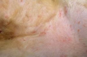
- Miliary dermatitis – small erythematous papules often covered with crusts. Many other skin disorders of cats can have miliary dermatitis. In fact, fleabite allergy is the most common cause of feline miliary dermatitis.
-
-
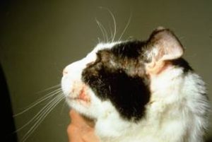
- Face, head and neck pruritus.
-
-
-
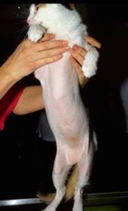
- Self-inflicted, non-inflammatory alopecia – Keep in mind that many other skin diseases in cats can have this clinical presentation. The other diseases include surface demodicosis caused by Demodex gatoi, food allergy, and psychogenic causes of self-inflicted alopecia.
-
-
-
- Lesions of the eosinophilic granuloma complex – Rule out atopic skin syndrome when cats are presented with lesions of the eosinophilic granuloma complex.
-

-
- The age of onset is variable ranging from 6 months to 8.5 years, with most cases below the age of 2 years.
- No sex or breed predilection has been reported.
- As in dogs, pruritus seems to be the hallmark of FASS and the most consistent sign. Be specific when investigating pruritus and ask owners if the cat is scratching, chewing/nibbling, rubbing and overgrooming. Owners may not report pruritus as cats often hide to itch. Therefore, do not rule out FASS if the owner does not notice the presence of pruritus. In this circumstance, perform a trichogram to determine if the tips of the hair shafts are broken-off indicating that the cat has caused the hair loss.
- Cats with FASS can exhibit their symptoms seasonally or perennially with most cats reportedly having year-round signs.
- Since pruritic cats can severely damage their skin, they are often presented to veterinary dermatologists early in the course of the disease, which can be difficult to determine seasonality.
- Secondary pyoderma and Malassezia dermatitis are not as common as in dogs. Nevertheless, check for their presence as they aggravate the allergic disease!
-
Differential diagnoses for the various clinical presentations of FASS
- Non-inflammatory alopecia:
- Parasitic: surface demodicosis, cheyletiellosis, flea infestation.
- Allergic: food allergy.
- Psychogenic.
- Endocrine: Cushing’s disease and hypothyroidism – very rare disorders in cats; moreover, most cases of non-inflammatory alopecia in cats are self-inflicted, therefore, not endocrine in origin. In addition, cats with Cushing’s disease and hypothyroidism have various systemic signs.
- Telogen effluxium.
- Miliary dermatitis:
- Infectious: bacterial, fungal.
- Ectoparasitic: demodicosis, cheyletiellosis, notoedric mange.
- Allergic: fleabite allergy (most common cause of miliary dermatitis in cats; be very suspicious if lesions are present on the caudal aspect of the body), food allergy, contact allergy.
- Eosinophilic granuloma complex:
- Food allergy, fleabite allergy.
- Non-inflammatory alopecia:
-
Diagnosis
- The diagnosis is usually based on:
- Compatible history and clinical signs.
- Exclusion of other pruritic skin diseases such as food allergy, pruritic parasitic diseases (e.g. surface demodicosis, cheyletiellosis), fleabite allergy and the other differentials listed above.
- Skin scrapings, tape preparations, impression smears, fecal flotations (cats ingest surface parasites when grooming), and treatment trials with parasiticides should be performed to rule out parasitic diseases.
- A strict food elimination trial with a home cooked or prescription novel limited ingredient or hydrolyzed protein diet should be performed after ruling out parasitic diseases in every case with nonseasonal pruritus.
- Cats with FASS should be placed on a parasiticide year-round, especially if residing in a geographic region where fleas are a problem.
- Allergen tests should be primarily performed to select allergens for allergen-specific immunotherapy, not to diagnose FASS.
- The diagnosis is usually based on:
-
Treatment
- Allergen-specific immunotherapy (ASIT) is the only treatment that modulates the allergic response. Discuss with owners the following before they commit to this treatment modality:
- False positive results can occur; therefore, it is important to select allergens for the ASIT that correspond to the cat’s history.
- Some cats will not react to any allergens included in the skin or serologic tests.
- Treatment can be administered via subcutaneous injections or sublingual route. The same protocol used for dogs is generally used for cats.
- Reported success rate is 50% to 75%. However, no placebo controlled trials have been performed to support this success rate.
- It may take 12 months before improvement can be seen.
- Cost of therapy per year.
- Glucocorticoids are very effective in treating FASS. The main concerning side effect in cats is diabetes mellitus and close monitoring is needed if using glucocorticoids long-term. Try to use oral and short acting drugs to minimize the risks for developing diabetes mellitus.
- Oral prednisolone not prednisone – prednisolone works better than prednisone because cats do not effectively convert prednisone to prednisolone, which is the active metabolite. The initial dose is 1.0 to 2.2 mg/kg q 24h until the patient is in remission (5 to 10 days). The dose should be reduced gradually until an alternate-day regimen with the lowest dose that controls the disease is achieved. Monitor for diabetes mellitus if used long-term.
- Oral triamcinolone acetonide – It is more potent than prednisolone, so only use it in cases that do not respond to prednisolone. The initial dose is 0.1 – 0.2 mg/kg q 24h until the patient is in remission (5-10 days). The maintenance dose is 0.05-0.1 mg/kg q 48h – 72h. Monitor for diabetes mellitus if used long-term.
- Oral dexamethasone – It is more diabetogenic and potent than prednisolone and triamcinolone, so only use oral dexamethasone if prednisolone and triamcinolone do not work. The initial dose is 0.125 – 1.0 mg/kg q 24h until the patient is in remission (5-10 days). The maintenance dose is the lowest effective dose that maintains the disease controlled and should be administered q 48h – 72h. Monitor for diabetes mellitus if used long-term.
- Methylprednisolone acetate (Depo-Medrol) injections should only be used when oral glucocorticoids cannot be used. The long-acting and more potent effects will increase the changes of inducing diabetes mellitus in cats. It is given intramuscularly at the dose of 4 to 5 mg/kg. Do not dose more frequently than every 8 weeks and preferably every 12 weeks. Do not administer more than three consecutive doses. Injectable glucocorticoids are not appropriate for long-term therapy due to the severe potential side effects associated with long-acting glucocorticoids (mainly diabetes mellitus in cats).
- Make sure to evaluate the cat’s heart condition before prescribing glucocorticoids to avoid heart failure, especially the long-acting formulations.
- Cyclosporine is effective in treating FASS.
- A liquid micro-emulsion formulation of cyclosporine containing 100mg/ml (Atopica®) is available for cats who need long-term therapy. Studies have shown that 7.0 mg/kg once daily is effective to manage cats with hypersensitivity dermatitis. It can be administered mixed in food or directly in the mouth. Not uncommonly, cats reject the liquid cyclosporine formulation.
- About 75% of cats respond to cyclosporine. Vomiting and diarrhea are the most common side effects. Maximal response is noted around 6 week after starting therapy, but response may be seen sooner. When significant results are noted, find the lowest possible dose regimen that maintains the disease under control; some cats can be maintained on 2-3 days per week dosing.
- Cats on cyclosporine should be kept indoors and not fed raw meat due to the risk of toxoplasmosis. Cats on long-term therapy should be monitored for gingival hyperplasia.
- We recommend having a chemistry profile and urinalysis performed every 6 to 12 months if a cat is to be maintained on long-term cyclosporine therapy.
- A study looked at the accuracy and precision of compounded cyclosporine capsules and solutions and found that the compounded cyclosporine solution may deviate by more than 10% from the labelled strength. The authors recommend that compounded cyclosporine only be prescribed when the commercial product cannot be used since the bioavailability and clinical efficacy of compounded cyclosporine is currently unknown.
- Oclacitinib (Apoquel®) has been shown to effectively manage cats with FASS. However, its use is off-label in cats.
- A pharmacokinetic study and clinical experience have shown that the effective dose in cats is 1 mg/kg q 12h to q 24h.
- Safety studies have shown it to be safe in cats but their duration was short. Therefore, because long-term safety data are lacking, it is recommended to monitor treated cats closely with a CBC, chemistry profile and urinalysis two to three times per year if using oclacitinib long-term.
- Antihistamines are typically used as adjunctive therapy.
- Perform a treatment trial to choose the best antihistamine to be used as maintenance therapy.
- The following antihistamines can be tried: chlorpheniramine (2-4 mg/cat q 12h), diphenhydramine (2.2 mg/kg q 12h), clemastine (0.68 mg/cat q 12h), hydroxyzine (10 mg/cat q 12h), cyproheptadine (2 mg/cat q 12h), amitriptyline (0.5-1 mg/kg q 12h) and cetirizine (5 mg/cat q 24 h). A common side effect associated with cyproheptadine is polyphagia.
- Omega-3 fatty acid supplements have been reported to be beneficial as adjunctive therapy in the management of cats with FASS.
- It will work best when given in association with antihistamines.
- The combined EPA and DHA dose can be 100 mg/kg q 24h.
- Ultramicronized palmitoylethanolamine at the dose of 10 mg/kg q 12h can be considered as an adjunctive therapy for cases that are not well controlled at low maintenance doses of immunomodulatories drugs such as glucocorticoids, cyclosporine and oclacitinib.
- Client education is the key for long-term successful management of FASS:
- Owners need to know that because the clinical presentation of FASS is quite variable and not specific, a step-by-step diagnostic approach taking in consideration the cat’s history and clinical signs will be needed to rule in/out other possible disorders.
- Owners need to know that it may take a while to find the best treatment regimen for the patient, which may entail various office visits.
- Remember to tell owners that FASS is an allergic disease and, as such, it cannot be cured but only controlled with continuous or intermittent use of medications.
- Allergen-specific immunotherapy (ASIT) is the only treatment that modulates the allergic response. Discuss with owners the following before they commit to this treatment modality:
Important Facts
- The pathogenesis of feline atopic skin syndrome (FASS) is not well understood.
- Clinical signs of FASS are quite variable and include four reaction patterns: military dermatitis, face-head-neck pruritus, self-inflicted non-inflammatory alopecia and lesions of the eosinophilic granuloma complex.
- Diagnosis is based on a compatible history, clinical signs and the exclusion of other pruritic dermatosis.
- The most important differential diagnoses are food allergy, fleabite allergy, pruritic parasitic skin diseases, and other causes of self-inflicted alopecia.
- A positive skin or serologic allergen test is not required for the diagnosis; however, it will identify the allergens for allergen-specific immunotherapy.
- Treatment options include allergen-specific immunotherapy, glucocorticoids, cyclosporine, oclacitinib, palmitoylethanolamine, antihistamines and fatty acid supplements.
- Monitor for side effects with a chemistry profile, urinalysis and urine culture at least once a year if the animal will be maintained on corticosteroids, cyclosporine or oclacitinib therapy.
References
Carrasco I, Ferrer L, Puigdemont A. Efficacy of oclacitinib for the control of feline atopic skin syndrome: correlating plasma concentrations with clinical response. J Feline Med Surg 2022; 24: 787-793.
Ferrer L, Carrasco I, Cristofol C et al. A pharmacokinetic study of oclacitinib maleate in six cats. Vet Dermatol 2019; DOI: 10.1111/vde.12819.
Foj R, Carrasco I, Clemente F, et al. Clinical efficacy of sublingual allergen-specific immunotherapy in 22 cats with atopic dermatitis. Vet Dermatol 2021; 32: 67–e12
Halliwell R, Pucheu-Haston CM, Olivry T et al. Feline allergic diseases: introduction and proposed nomenclature. Vet Derm 2021; DOI: 10.1111/vde.12899.
Hobi S, Linek M, Marignac G et al. Clinical characteristics and causes of pruritus in cats: a multicentre study on feline hypersensitivity-associated dermatoses. Vet Dermatol 2011; 22:406-413.
King S et al. A randomized double-blinded placebo-controlled study to evaluate an effective ciclosporin dose for the treatment of feline hypersensitivity dermatitis. Vet Dermatol 2012; 23: 440-447.
Lopes, NL, Campos DR, Machado MA, et al. A blinded, randomized, placebo-controlled trial of the safety of oclacitinib in cats. BMC Vet Res 2019; 15: 137.
Miller, Griffin, Campbell. Muller and Kirk’s Small Animal Dermatology. 7th Edition. In: Hypersensitivity Disorders. Elsevier Health Sciences. 2013 p 388–392.
Mueller RS, Nuttall T, Prost C et al. Treatment of the feline atopic syndrome – a systematic review. Vet Derm 2021; DOI: 10.1111/vde.12933.
Noli C, Matricoti I, Schievano A. A double-blinded, randomized, methylprednisolone-controlled study on the effecicay of oclacitinib in the management of pruritus in cats with nonflea nonfood-induced hypersensitivity dermatitis. Vet Derm 2021; 30:110-e130.
Noli C, della Valle MF, Miolo A et al. Effect of dietary supplementation with ultramicronized palmitoylethanolamide in maintaining remission in cats with nonflea nonfood-induced hypersensitivity dermatitis: a double-blind, multicentre, randomized, placebo-controlled study. Vet Derm 2019;; 30: 387-e117.
Ployngam T, Tobias A, Smith S et al. Hemodynamic effects of methylprednisolone acetate administration in cats. Am J Vet Res 2006; 67:583-587.
Reedy, Miller, Willemse: Urticaria, Angioedema, and Atopy. In: Allergic Skin Diseases of Dogs and Cats, 2nd ed, W.B. Saunders, Philadelphia, PA, 1997, p 44 – 45.
Roberts ES, Speranza C, Friberg C et al. Confirmatory field study for the evaluation of ciclosporin at a target dose of 7.0 mg/kg (3.2 mg/lb) in the control of feline hypersensitivity dermatitides. J Fel Med Surg 2016; DOI:10.1177/1098612X16636660.
Santoro D, Pucheu-Haston CM, Prost C et al. Clinical signs and diagnosis of feline atopic syndrome: detailed guidelines for a correct diagnosis. Vet Derm 2021; DOI: 10.1111/vde.12935.
