2.3 Atopic Dermatitis – Horses
Learning Objectives
- Learn how atopic dermatitis manifest clinically in horses
- Learn how to diagnose atopic dermatitis in horses
- Learn the treatment options for atopic dermatitis in horses
-
General Considerations
- Atopic dermatitis is an inherited predisposition to produce increased amounts of sensitizing antibodies to common environmental antigens that in non-atopic individuals cause no disease. Examples of such allergens include pollens of grasses, weeds, and trees, molds, house dust and storage mites and barn dust.
- It is known that IgE reaginic antibodies and type I hypersensitivity reactions occur in horses, supporting the belief that they are capable of developing atopic dermatitis.
-
Pathogenesis
- Like in other species, the pathomechanism of atopic dermatitis is not well understood in horses.
- Part of the mechanism involves IgE sensitizing antibodies. These antibodies bind to high affinity receptors on skin mast cells and when a specific allergen binds to two adjacent antibodies, it cross-links the IgE molecules resulting in mast cell release of inflammatory mediators.
- Release of mast cell mediators results in a cascade of events that culminate in pruritus and inflammation.
- Similar to dogs and humans, Langerhans’ cells in the epidermis and T cells (Th2) in the dermis are also likely to play a role in disease development. In fact, Th2 cells predominance, as demonstrated in dogs and humans, leads to increased production of allergen-specific IgE antibodies. Moreover, skin barrier defect as well as genetic and environmental factors may also be involved in the pathogenesis.
-
Clinical Signs
- Clinical signs most commonly begin between 1 ½ and 6 years of age.
- Anecdotal reports indicate that equine atopic dermatitis can have familial and breed-related predispositions. Thoroughbreds and Arabians (and perhaps Quarter Horses, Warmbloods, and Morgans) may be at risk. However, more evidence is needed to confirm or refute these facts.
- The major clinical sign of equine atopic dermatitis is pruritus or urticaria associated or not with pruritus.
- Pruritus may be seasonal or year-round corresponding to the allergens that trigger the hypersensitivity reaction.
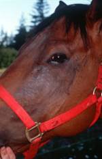
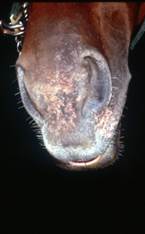
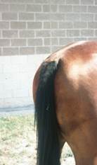
- Pruritus and secondary self-inflicted lesions of alopecia and excoriation typically occur on the face, ears, ventrum and legs. As he disease becomes chronic, lichenification, hyperpigmentation and hypopigmentation develop.
-
- Clinical signs may resemble insect bite hypersensitivity.
- In some horses, pruritus may be accentuated in areas not typical for insect hypersensitivity, making differentiation of the two disorders easier.
- Some atopic horses have concurrent insect-bite hypersensitivity and/or food allergy.
- Secondary bacterial folliculitis may occur, which can aggravate the clinical signs of atopic dermatitis.
- There is conflicting evidence about the role of atopy in recurrent airway obstruction (RAO) formerly known as obstructive pulmonary disease [COPD].
- A subset of horses with RAO seems to be truly atopic, based on intradermal testing, response to allergen challenge, and response to allergen-specific immunotherapy.
-
Diagnosis
- Diagnosis is based on a compatible history, characteristic clinical signs, and ruling out other pruritic dermatoses (e.g. food allergy, insect bite hypersensitivity, lice (pediculosis), pruritic mange).
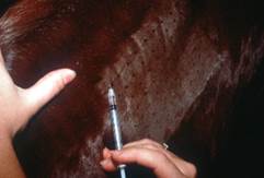
- Intradermal testing will identify the allergens that the horse has become sensitized. The main value of this test is to select antigens for the allergen-specific immunotherapy vaccine. This should not be used as a diagnostic test since false positive and false negative reactions occur.
-
Treatment
- Allergen-specific immunotherapy (ASIT):
- The only treatment that can modify the allergic response is allergen-specific immunotherapy (ASIT) based on intradermal and serum allergen-specific IgE tests.
- Intradermal allergen test requires the discontinuation of antihistamines for at least 7 days before testing. Ideally, glucocorticoids should be withdrawn at least 14 days before testing.
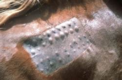
- The same technique described for testing dogs can be used in horses. Fractious animals can be sedated with xylazine hydrochloride. The skin over the lateral neck is the preferred test site. Gently, clip the hair with a No. 40 blade using no chemical preparation to clean the test site.
- Read test results at 15’, 30’, 4 and 6 hours after finishing the test. Delayed reactions appear to be present in horses. However, a study showed that little additional information is gained with the 24-hours assessment because few allergens will have a positive reaction at 24-hours without previous reaction at 15’, 30’, and 4 to 6 hours.
- Remember! A positive skin or serum test reaction tells us that the horse can make allergen-specific IgE; it does not indicate that the antigen in question participates in disease development. In other words, false-positive reactions and false-negative reactions can occur with intradermal and serum testing. Therefore, the results of these tests must correlate with the disease seasonality and environment of the horse to increase the chances of including in the vaccine allergens that are clinically relevant to the patient. For example, if a horse is not pruritic at the time a plant pollinates but has an intradermal or serological test reaction to that plant’s pollen, this allergen should not be included in the immunotherapy.
- ASIT based on serum allergen-specific IgE has been used more often in horses with atopic dermatitis. One of the reasons for large numbers of false-positive reactions in serum allergen-specific IgE tests is the presence of IgE antibodies against cross-reactive carbohydrate determinants (CCD). Plants and insects express these glycoproteins. Some laboratories developed an assay that inhibits the IgE antibodies against CCD. This could improve the specificity of the serum allergen-specific IgE test and ultimately improve the outcome of immunotherapy based on serum test results.
- A study demonstrated that there are no significant differences in the ASIT response regardless if the intradermal or serum allergen-specific test is used for the selection of allergens to compound the immunotherapy. Eighty four percent of the horse owners in this study reported that immunotherapy helped improve the clinical signs of their horses with atopic dermatitis and 59% of these horses only required this treatment modality to control their disease.
- Another study of horses with atopic dermatitis treated with ASIT showed an improvement in clinical signs of 76.5% after 12 months of therapy and of 82% after 24 months of therapy.
- Horse owners have to understand that it may take 12 months before any benefit from immunotherapy can be noted and that the overall success rate is about 60-80%.
- Glucocorticoids:
- Symptomatic therapy with glucocorticoids is often used to control pruritus and inflammation in horses with atopic dermatitis. Prednisolone and dexamethasone are the glucocorticoids used more often to manage equine pruritic dermatoses.
- Oral prednisolone: Prednisone is not effective in horses because of poor absorption, rapid elimination, and failure of hepatic conversion of prednisone to prednisolone, the active metabolite. Therefore, avoid using prednisone in horses. Induction doses of prednisolone (0.5 – 2.0 mg/kg q 24h) are needed until remission is achieved and then a tapering dose regimen is attempted to arrive at a safe alternate-day maintenance dose (0.25 to 0.5 mg/kg q 48h). Most induction periods range from 7 to 14 days, followed by a tapering period of 2 to 5 weeks.
- Oral dexamethasone: Dexamethasone is more potent than prednisolone and can be tried in horses that do not respond to prednisolone. In many cases, an induction dose of 0.02 to 0.1 mg/kg q 24h can produce remission in a few days. Thereafter, taper the dose to 0.01 to 0.02 mg/kg q 48h to 72h as maintenance therapy.
- Adverse effects: The most concerning side effects more likely to occur with long-term therapy, include immunosuppression, delayed wound healing, and insulin-resistant laminitis. Various studies have evaluated the risk of developing laminitis in horses treated with glucocorticoids and most have been unable to show an association. However, horses with metabolic syndrome or Cushing’s disease are at a higher risk. If glucocorticoids are needed in these patients, avoid triamcinolone and use prednisolone instead, as the duration of insulin resistance is longer with triamcinolone, intermediate with dexamethasone and shorter with prednisolone. Other adverse effects include muscle wasting, hyperglycemia, polyuria and polydipsia, and rarely gastric ulcer and hepatic lipidosis.
- Antihistamines:
- Antihistamines have fewer side effects than glucocorticoids and provide an alternative for horses with mild disease or that cannot tolerate glucocorticoids. However, they may not be effective as sole therapy for horses with moderate or severe atopic dermatitis but can be used as adjunctive therapy to glucocorticoids.
- Cetirizine (0.2–0.4 mg/kg PO q 12h) and hydroxyzine hydrochloride/pamoate (1–2 mg/kg q 8–12 h) are generally the preferred antihistamines to manage both urticaria and allergic hypersensitivities in horses. It is generally more effective for urticaria than for pruritus.
- Other less effective antihistamines include diphenhydramine (1-2 mg/kg q 8h – 12h), doxepin hydrochloride (0.75 to 1.0 mg/kg q 12h), and chlorpheniramine (0.25-0.5 mg/kg q 12h).
- The only side effects observed with antihistamines are occasional episodes of sedation and personality changes.
- Randomized, placebo-controlled clinical trials are needed to investigate the efficacy of antihistamines in horses with atopic dermatitis.
- Pentoxifylline:
- Pentoxifylline is a phosphodiesterase inhibitor that has multiple immunomodulatory properties and minimal side effects (hyperexcitability, sweats).
- The proposed dose is 10–15 mg/kg q 12h but the exact therapeutic dose is not currently known. To increase efficacy consider increasing the dosing frequency to three times daily.
- It can be used in conjunction with glucocorticoids with the purpose of having a synergistic effect or of tapering the glucocorticoid dose. However, there is no randomized, controlled clinical trials to support its use in horses with atopic dermatitis.
- Essential fatty acids:
- Currently there is very little evidence to support the use of essential fatty acids to treat allergic diseases in horses.
- One randomized controlled clinical trial showed a decrease in intradermal test reactions after 42 days of flax seed supplementation (omega 3 fatty acid; 1 lb/1000 lb/ kg q 24h) in horses with Culicoides hypersensitivity compared to controls.
- In an open clinical trial, five of 14 horses with insect bite hypersensitivity showed a very good response and five of 14 showed a good response after 13 weeks supplementation with 20g of evening primrose oil (omega 6) and marine fish oil (omega-3, 80:20 mixture).
- Oclacitinib (Apoquel):
- Two pharmacokinetic studies have shown that the half-life of oclacitinib in horses is longer than that in dogs. One study investigated a single oral dose of 0.2 mg/kg and the half-life was 9h to 10h. Another study used a single oral dose of 0.5 mg/kg and the half-life was similar ranging from 7h to 8h. The results of these studies suggest that oclacitinib can be used once daily in horses.
- The efficacy and safety of two oclacitinib doses (0.1 mg/kg and 0.25 mg/kg q 24h) in horses with allergic diseases were compared to placebo in a randomized trial of 28 days. Pruritus (PVAS) and lesion scores were assessed. No significant differences in lesion scores were noted between the groups. Starting on day 5 until day 28, a significant treatment difference in PVAS scores was noted in horses treated with oclacitinib at 0.25 mg/kg q 24h compared to placebo. Clinical pathology findings showed no effects that appeared to be clinically or biologically significant.
- More controlled clinical trials are needed to determine the efficacy of oclacitinib in treating equine atopic dermatitis, the effective dose range, and the long term safety profile.
- Topical therapy:
- Medicated shampoos can be used as adjunctive therapy because they remove surface irritants, bacteria and allergens. In addition, bathing hydrates the skin and improves the integrity of the epidermal barrier. Horses should be bathed in cool water because it results in vasoconstriction hence decreasing delivery of inflammatory mediators to the skin.
- Topical therapy containing antiseptics such as, chlorhexidine, benzoyl peroxide, hypochlorous acid, and essential oils in the form of mousse, spray or rinse is a good adjunctive measure to treat or prevent secondary bacterial and fungal infections. They have the advantage of leaving a residual effect and are often used in between bathing.
- Avoidance:
- Unfortunately, avoidance of aeroallergens is frequently impossible or impractical. Nevertheless, any effort to reduce the allergen load may be beneficial.
- Because atopic dermatitis can co-exist with insect bite hypersensitivity, strict insect avoidance will increase the pruritus and inflammation thresholds and optimize response to therapy.
- Allergen-specific immunotherapy (ASIT):
-
Prognosis and client education
- Atopic dermatitis has a good prognosis if managed early in the disease course.
- Horse owners have to understand the following:
- The allergic tendency is an inherited trait; however, the specific genetic mechanisms have not been elucidated in the horse.
- Atopic dermatitis is a manageable condition, not a curable disease.
- Each case is unique and the ultimate goal is to delineate the best treatment plan for the patient, which takes time and may entail various visits.
Important Facts
- More studies need to be conducted to better understand the pathomechanism of atopic dermatitis in horses, and ultimately investigate more effective and safe treatment modalities.
- Pruritus can be seasonal or year-round correlating with the environmental allergens that trigger the hypersensitivity reaction.
- Pruritus and self-inflicted lesions of alopecia and excoriation are often present on the face, ears, ventrum and legs. Hyperpigmentation, hypopigmentation and lichenification develop as the disease becomes chronic.
- Clinical signs can mimic insect hypersensitivity and atopic dermatitis and insect bite hypersensitivity can co-exist.
- Diagnosis is based on a compatible history, characteristic clinical signs and exclusion of other pruritic dermatoses.
- False positive and false negative results can occur with the intradermal and serum allergen tests; therefore, they cannot be used to confirm the diagnosis of atopic dermatitis but should be used to select allergens for the allergen-specific immunotherapy.
- Allergen selection for immunotherapy has to correlate with disease seasonality and the horse environment to increase the chances of success.
- Treatment should be tailored to each individual horse and includes the following: allergen-specific immunotherapy; glucocorticoids, antihistamines (often adjunctive), pentoxifylline (often adjunctive), and topical therapy. These treatment options are often combined to achieve the best results.
References
Costa LRR, Johnson JR, Baur ME, et al. Temporal clinical exacerbation of summer pasture associated recurrent airway obstruction and relationship with climate and aeroallergens in horses. Am J Vet Res 2006; 67(9):1635–1642.
Enck K, Lee K, McKinney B, Blankenship KD, Montesano C. Detection and inhibition of IgE antibodies reactive with cross-reactive carbohydrate determinants in an ELISA for allergen specific IgE in horses. Vet Dermatol. 2020;32:685–e184.
Fadok VA. Update on equine allergies. Vet Clin Equine 2013; 29: 541–550.
Fadok, V.A. Overview of Equine Pruritus. In: Veterinary Clinics of North America: Equine Practice, W.B. Saunders, Philadelphia, PA, 1995, p 1-10.
Felippe M. Julia B. Immunosuppressive therapy in Equine Clinical Immunology; Felippe M. Julia B., Wiley-Blackwell Ames Iowa 2016; 237-238.
Friberg CA, Logas D. Treatment of Culicoides hypersensitive horses with high-dose n-3 fatty acids: a double-blinded crossover study. Vet Dermatol 1999; 10: 117–122.
Jose-Cunilleras E, Kohn CW, Hillier A et al. Intradermal testing in healthy horses and horses with chronic obstructive pulmonary disease, recurrent urticaria, or allergic dermatitis. J Am Vet Med Assoc 2001; 219(8): 1115-1121.
Hopke K, Coleman M, Kneese E et al. Prevalence of serum immunoglobulin E against cross-reactive carbohydrate determinants in allergic and nonallergic horses, and its impact on polysensitisation in serum allergen tests. Vet Derm 2022; DOI: 10.1111/vde.13073.
Leclere M. Corticosteroids and immune suppressive therapies in horses. Vet Clin North Am Equine Pract 2017; 33:17-27.
Lorch G, Hillier A, Kwochka KW et al. Comparison of immediate intradermal test reactivity with serum IgE quantitation by use of a radioallergosorbent test and two ELISA in horses with and without atopy. J Am Vet Med Assoc 2001; 218(8):1314-1322.
Marsella R. Pruritic horse: Approach to allergic skin diseases in horses. Vet Clin Equine 2024; 40:219-235.
Marsella R, White S. Fadok VA, et al. Equine allergic skin diseases: Clinical consensus guidelines of the World Association for Veterinary Dermatology. Vet Derm 2023; 34: 175-208.
Marsella R. Equine allergy therapy: Update on the treatment of environmental, insect bite hypersensitivity, and food allergy. Vet Clin Equine 2013; 29: 551-557.
O’Neill W, McKee S, Clarke AF. Flaxseed (Linum usitatissimum) supplementation associated with reduced skin test lesional area in horses with Culicoides hypersensitivity. Can J Vet Res 2002; 66: 272–277.
Petersen A, Schott HC. Effects of dexamethasone and hydroxyzine treatment on intradermal testing and allergen-specific IgE serum testing results in horses. Vet Dermatol
2009; 20:615–22.
Radwanski NE, Morris DO, Boston RC et al. Longitudinal evaluation of immunological responses to allerge-specific immunotherapy in horses with IgE associated dermatological disease, a pilot study. Vet Derm 2019; DOI: 10.1111/vde.12732
Rashmir-Raven A. Equine Dermatology: Allergic and Immune Mediated Dermatitis – The Diagnosis and Treatment. In Proceedings of the Pacific Veterinary Conference: São Francisco, CA, USA: 2016.
Scott, D.W.. Immunologic Diseases. In: Large Animal Dermatology. 1st ed., W.B. Saunders, Philadelphia, PA, 1988, p 294-300.
Stepnik CT, Outerbridge CA, White SD et al. Equine atopic skin disease and response to allergen-specific immunotherapy: a retrospective study at the University of California-Davis (1991-2008). Vet Derm 2012; 23, 29–e7
Visser M, Cleaver D, Cundiff B, King V, Sture G. Oclacitinib maleate (Apoquel) dose determination in horses with naturally occurring allergic dermatitis. 2020 ACVIM forum on demand research abstract program. J Vet Int Med 2020; 34: 2977–2978.
Volland-Francqueville M, Sabbah A. Recurrent or chronic urticaria in Thoroughbred racehorses: clinical observations. Allerg Immunol (Paris). 2004;36(1):9–12.
Yu AA. Equine Allergies: Treatment of Equine Allergies. In Proceedings of the Ontario VMA Conference & Trade Show : Ontario, VMA, USA: 2016.
