2.4 Adverse Food Reaction/Food Allergy – Dogs and Cats
Learning Objectives
- List the three most common causes of food allergy in dogs and cats.
- Know the characteristic history and clinical signs of food allergy in dogs and cats.
- List the steps involved in doing an elimination diet trial and how to interpret the results.
- Know the pros and cons of the various diets used in food elimination trials.
- Know the important client education points.
- Know the important points you have to convey to clients before starting a food elimination trial.
- Know the important points you have to convey to clients regarding the long-term management of a pet with food allergy.
-
General Considerations
- An adverse reaction to dietary ingredients includes two categories: (i) immunologically mediated or allergic reaction and, (ii) non-immunologically mediated reaction or food intolerance. An example of non-immunologically mediated reaction is the presence of vasoactive amines in foods such as histamine in scombroid fish including mackerel, tuna, mahi-mahi, and sardines. Depending on the amount present, these vasoactive amines can lead to clinical signs that may mimic food allergy.
- The cutaneous signs of food intolerance are quite variable and, not always, the dog and cat are pruritic.
- In this chapter, we will discuss the immunologically mediated component of adverse food reaction, thus, hypersensitivity reactions to food allergens or food allergy.
- Clinical signs of food allergy may include cutaneous and gastrointestinal signs but the latter is more frequently seen with food intolerance.
- The clinical signs of food allergy in most dogs and cats are indistinguishable from those of atopic dermatitis because food allergens trigger the atopic disease.
- Food allergy is a nonseasonal pruritic dermatosis of dogs and cats.
- In a systematic review paper, estimates of food allergy prevalence among dogs with skin diseases ranged from 0 to 24% and among cats with skin diseases from 3 % to 13%. The heterogeneity of data can be explained by differences in the geographical populations investigated, diversity in the groups of animals included in the studies, how systematically food allergy was investigated in the regions included, and the method used to diagnose food allergy.
-
Cause and Pathogenesis
- The pathogenesis of food allergy in dogs and cats is still not well understood but is likely similar to that of atopic dermatitis as the clinical signs of these two diseases are indistinguishable.
- It typically occurs after the pet has been on the same diet for months to years. When owners report that the clinical signs developed after changing to a new diet, the protein source is the same in many cases.
- Most common allergens – The information below was retrieved from a 2016 publication on “Critically appraised topics on adverse food reaction of companion animals: common food allergens in dogs and cats”. The caveat is that these data came from Australia, Europe and North America and may only be applicable to these geographical regions. Knowing the most common food allergens of pets in a region can be valuable in choosing the foods to include in a provocation test when an accurate pet’s dietary history is impossible or difficult to obtain.
- Dogs:
- Beef (34%), dairy products (17%), chicken (15%), wheat (13%), lamb (5%).
- Cats:
- Beef (18%), fish (17%), chicken (5%).
- Dogs:
-
Clinical Signs
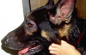
- Canine:
- In food allergy or food-induced atopic dermatitis, the pruritus is year-round because the animal is typically exposed to the food allergen(s) in the diet throughout the year. In contrast, in environmental-induced atopic dermatitis the pruritus can be seasonal or year-round.
- As mentioned previously, atopic dermatitis and food allergy can look the same and cannot be differentiated based on clinical signs. In this context and if food is identified as the trigger allergen, the disease is called “food-induced atopic dermatitis”. You most likely will see these cases in practice. It is important, therefore, to perform a strict food elimination trial in every dog with nonseasonal signs of atopic dermatitis to determine if food allergens are contributing to the clinical signs.
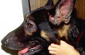
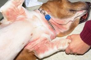
- Body areas typically affected mimic that of environmental-induce atopic dermatitis.
- Face, feet, axilla, groin
- Canine:
-
-
-
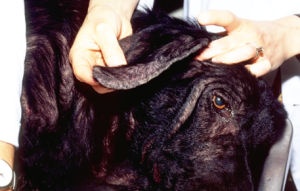
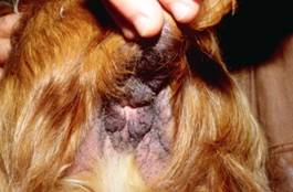
- “Ears and Rears” – recurrent otitis and perianal dermatitis.
- Food-induced and environmental-induced atopic dermatitis can rarely mimic scabies and fleabite allergy but these diseases have to be included in the list of differential diagnoses.
-
-
-
-
-
- Gastrointestinal signs may be seen in conjunction with dermatologic signs in about 10-40% of the cases. They include vomiting, diarrhea, colic, excessive flatulence, and more than three bowel movements per day.
- What lesions do we see? Lesions will mimic those of environmental-induced atopic dermatitis.
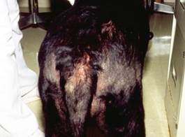
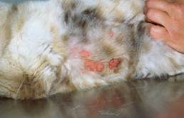
- Lesions secondary to self-trauma from pruritus: alopecia, excoriations, acute moist dermatitis.
-
-
-
-
-
- Lesions secondary to chronic inflammation: lichenification and hyperpigmentation.
-
-
-
-
-
- Lesions associated with secondary staphylococcal pyoderma, which can present as papules, pustules, crusts, moth-eaten alopecia and epidermal collarettes.
- Lesions associated with Malassezia dermatitis, which can present as erythema, scales, brownish discoloration of skin and hair, lichenification.
- Lesions associated with secondary seborrhea, which will present as scales with or without excessive sebum production.
- Age:
- It can occur before 1 year of age to more than 10 years of age. However, most cases will develop signs between 1 to 3 years of age.
- A fair number of food-induced atopic dermatitis occurs in dogs before 1 year of age and should raise the index of suspicion for this disease above that of environmental-induced atopic disease.
-
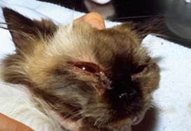
- Cats:
- In food allergy, the pruritus is always year-round in contrast with feline atopic skin syndrome (FASS) that it can also be seasonal. However, the clinical signs of food allergy in cats are similar to the signs of FASS.
- Where are they pruritic?
- Face
-
-
-
-
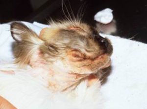
- Head and neck
-
-
-
-
-
- Generalized
- Pruritic otitis externa.
- What lesions do we see? Similar to FASS the clinical signs are variable and mimic many dermatoses.
- Miliary dermatitis (widespread papulocrustous dermatitis). Many other skin disorders of cats can have miliary dermatitis. These include FASS, fleabite allergy, (most common), contact allergy, demodicosis, cheyletiellosis, notoedric mange, bacterial and fungal infections.
-
-
-
-
-
- Self-inflicted non-inflammatory alopecia. Keep in mind that many other skin diseases in cats can have this clinical presentation. The other diseases include FASS, surface demodicosis caused by Demodex gatoi, and psychogenic causes of self-inflicted alopecia.
-
-


-
-
-
- Lesions of the eosinophilic granuloma complex. FASS can also manifest with lesions of the eosinophilic granuloma complex.
-
-
-
-
-
- Gastrointestinal signs are uncommon.
-
-
-
Diagnosis
- Characteristic history (year-round pruritus) and clinical signs (mimic environmental-induced atopic dermatitis in dogs and FASS in cats) are important parts of the diagnostic equation.
- The only reliable method of testing for food allergy is a dietary elimination and challenge trials. The intradermal and/or serum allergen tests are not reliable in diagnosing food allergy.
- Dietary elimination and challenge trials:
- The crucial part of the food elimination trial is to select a proper diet. The options are to select a novel protein and carbohydrate diet, a hydrolyzed protein diet or a diet where synthetic amino acids are the source of protein (elemental diet).
- A diet with novel sources of protein and carbohydrate is selected based on a thorough dietary history. However, this diet can be quite difficult to find because pet owners often change diets, give table foods and will not remember all foods that the pet has ingested.
- Another potential issue in using a novel protein diet is the increasing concern about cross-reactivity between allergens of close (i.e. phylogenetically related) or distant animal species used as food sources. Examples of cross-reactivity between phylogenetically related species are beef and venison and chicken and duck. Examples of cross-reactivity between phylogenetically distant species are chicken and fish and beef and sheep. The clinical relevance of these findings is not known at this time. However, it is possible that the novel protein in the trial diet is duck but the dog has eaten chicken in which case the trial will be unsuccessful.
- A hydrolyzed or elemental diet is a good option when accurate dietary histories cannot be obtained. The molecular weight of the protein (carbohydrates are not hydrolyzed) in hydrolyzed diets is considered too small to be antigenic (<5 kDa). Ideally, the protein molecular weight should be <1 KDa (ultra-hydrolyzed). However, not all proteins in these diets are hydrolyzed, which can be a problem. Therefore, if possible choose an ultra-hydrolyzed diet where the protein source is novel. Another option that in theory eliminates these issues is to choose an elemental diet where synthetic amino acids are incorporated into the diet as the protein source. However, an elemental diet is currently very expensive.
- Homemade or commercial diets can be used and there are advantages and disadvantages to each one.
- Homemade diets with novel source of protein and carbohydrate:
- Advantages:
- No additives or preservatives.
- One source of protein and carbohydrate.
- You know each ingredient in the diet.
- Disadvantages:
- Not nutritionally balanced.
- Time consuming to prepare.
- It can be difficult to select a novel source of protein and carbohydrate.
- Advantages:
- Commercial diets – novel protein and carbohydrate diet, hydrolyzed protein diet, elemental diet.
- Advantages:
- Easy to feed.
- Nutritionally balanced.
- Preferred in animals less than 10 months old.
- Disadvantages:
- More than one source of protein and/or carbohydrate.
- Diet with novel sources of protein and carbohydrate can be difficult to find.
- Not all proteins in the diet are hydrolyzed.
- Contain additives and preservatives.
- Contamination with other allergens during processing.
- Not all ingredients are included in the label.
- Advantages:
- An important thing to keep in mind when using commercial diets is that they can be contaminated with storage mites, if not stored properly. The problem is the extensive cross reactivity that exists between house dust mites and storage mites. Consequently, atopic dogs and cats sensitized to house dust mites will likely have a positive challenge response if the original diet is contaminated with storage mites, which could result in an erroneous diagnosis of food allergy. The recommendation is to buy a new bag of the original diet to be used during the challenge part of the trial. In addition, it is best to keep commercial dry dog foods indoors and in temperate conditions and to seal the bags to decrease the risk of their contamination with storage mites.
- Dogs and cats less than 10 months old should be supplemented with vitamins and calcium during an elimination trial with homemade diet.
- Cats should be supplemented with taurine if a homemade diet is used during the trial.
- The food elimination trial is constituted of three parts: 1) food elimination; 2) food challenge; 3) food provocation.
- Food elimination trial:
- Introduce the selected trial diet gradually to prevent gastrointestinal signs – start by mixing only 25% of the trial diet and increase the amount daily by 25% so that in the fourth day the pet will be eating exclusively the trial diet. In cats or finicky dogs, the transition may need to be slower.
- The selected trial diet should be given exclusively for 8 weeks or until a significant decrease (at least 50%) in pruritus level is noted by the owner.
- If the animals are very itchy at the start of the trial and no secondary skin infection is present, it is reasonable to start the patient on an anti-pruritic and anti-inflammatory drug (e.g. glucocorticoid, oclacitinib). However, discontinue the medication at mid-trial to assess response. If the signs do not return, challenge the pet with the previous diet(s). If the signs return, put the pet again on the anti-pruritic and anti-inflammatory drug and continue the trial. At the end of the trial, discontinue the drug and assess the itch level again.
- At least 50% decrease in pruritus level indicates that food allergy is at least part of the problem; however, a challenge trial should always be performed to confirm a presumptive diagnosis of food allergy.
- Food allergy can occur with atopic dermatitis or fleabite allergy and this explains the partial response to a strict trial seen in some cases.
- Food challenge trial:
- It is used to confirm that the observed decrease in pruritus during the food elimination trial was due to food allergy and not a variation in environmental allergens or other reasons.
- The challenge consists in feeding the animal its regular diet and seeing the pruritus recur or get worse within 14 days. Usually, signs recur or worsen after 3 to 7 days of challenging but it may take up to 14 days for the signs to recur. In some cases, it may be noticed after only 3 to 72 hours post-challenging. If the pruritus recurs at any time during the challenge period, the dog should be placed again on the trial diet and the clinical signs should improve if the pet truly has food allergy. At this time, you can discuss with the owner the option of doing a provocation test.
- Food provocation trial:
- It is used to determine the food items to which the animal is allergic.
- A single food item is added to the elimination trial diet for up to 14 days.
- If the pruritus recurs or gets worse, remove the added food item and wait for the clinical signs to improve before testing another food item.
- The number of food items to be tested should be based on the ingredients in the culprit diet and decided by the pet owner and attending veterinarian.
- The provocation test can be very time consuming
- Educating the client:
- Client education is crucial for a successful food trial.
- Clients have to understand that if the trial is not strict and performed properly, there is no value in investing the time and money to doing it.
- Investigate carefully if the client’s household has the conditions to support a strict food elimination trail.
- All family members have to be involved for the trial to be successful.
- Be sure the owner discontinues any chews, treats, table scraps, vitamins, chewable heartworm, fatty acids, and medication in food. Non-flavored nylabones, rope chews, and rubber toys are fine.
- Advise owners to buy a new bag of the original diet to use during the challenge phase of the trial. This will prevent an erroneous diagnosis, which could occur if the old bag of food was contaminated with storage mites and the dog was sensitized to house dust mites. It is always important to keep bags of dry commercial foods in temperate conditions inside the house and sealed to avoid contamination with storage mites.
- Food elimination trial:
-
Treatment
- Be sure to identify and treat any secondary problems such as staphylococcal pyoderma, seborrhea and/or Malassezia dermatitis.
- Feed the diet that the pet tolerates exclusively throughout life.
- Because novel protein/carbohydrate diets and some hydrolyzed diets are not truly hypoallergenic, the animals may eventually develop allergy to the ingredients present in the maintenance diet. However, this happens rarely.
- Client education is the key for long-term successful management of food allergy.
- Food allergy is manageable but not curable.
- Feed exclusively the diet that the pet tolerates throughout the pet’s life.
- A provocation test can be done to determine the specific food items that the pet can or cannot eat.
- Food allergy can co-exist with atopic dermatitis or fleabite allergy and identifying and addressing any concurrent allergy will improve the long-term management of food allergy.
Important Facts
- Food allergy is associated with year-round symptoms.
- Pruritus is the hallmark of food allergy.
- Food antigens can trigger signs of atopic dermatitis; therefore, food allergy and atopic dermatitis cannot be differentiated based on clinical signs. In other words, atopic dermatitis can be induced by food or environmental allergens.
- Similarly, in cats, the clinical signs of food allergy and FASS are similar.
- Intradermal and serologic tests are not reliable diagnostic tests for food allergy.
- Elimination diet and challenge trials are the only reliable methods of testing for food allergy.
- Make sure to select the diet to be used in the trial very carefully. This can be a challenge!
- Options of diets include a single and novel protein and carbohydrate diet (commercial or homemade), a hydrolyzed-protein diet, or an elemental diet.
- The trial diet has to be given, exclusively, for 8 weeks or until significant improvement is noticed, whichever comes first.
- A decrease in pruritus of at least 50% is considered a positive test.
- Diagnosis is confirmed by feeding the animal its regular diet and seeing the pruritus recur within 14 days. The pet’s clinical signs have to improve again when the trial diet is reinstituted before confirming the diagnose.
- Be sure to identify and treat any secondary problem such as staphylococcal pyoderma, Malassezia dermatitis and seborrhea.
- Client education is crucial for conducting a success diet trial and for the long-term management of food allergy.
References
Jackson HA. Food allergy in dogs and cats; current perspectives on etiology, diagnosis, and management. J Am Vet Med Assoc 2023; doi.org/10.2460/javma.22.12.0548.
Miller, Griffin, Campbell. Muller and Kirk’s Small Animal Dermatology. 7th Edition. In: Hypersensitivity Disorders. Elsevier Health Sciences. 2013 p 400-405.
Mueller R, Olivry T, Prelaud P. Criticalyl appraised topic on adverse food reaction of companion animals (2): common food allergen sources in dogs and cats. BMC Vet Res 2016; DOI 10.1186/s12917-016-0633-8.
Olivry T, Meller R, Prelaud P. Criticalyl appraised topic on adverse food reaction of companion animals (1): duration of elimination diets. BMC Vet Res 2015; DOI 10.1186/s12917-015-0541-3.
Olivry T, Mueller RS. Critically appraised topic on adverse food reactions of companion animals (3): prevalence of cutaneous adverse food reactions in dogs and cats. BMC Vet Res 2017; DOI 10.1186/s12917-017-0973-z.
Olivry T, Mueller RS. Critically appraised topic on adverse food reactions of companion animals (7): signalment and cutaneous manifestations of dogs and cats with adverse food reactions. BMC Vet Res 2019; doi.org/10.1186/s12917-019-1880-2.
Olivry T et al. Critically appraised topic on adverse food reactions of companion animals (8): Storage mites in commercial pet foods. BMC Vet Res 2019; !5:385.
Olivry T, Mueller RS. Critically appraised topic on adverse food reactions of companion animals (9): time to flare of cutaneous signs after a dietary challenge in dogs and cats with food allergies. BMC Vet Res 2020; doi.org/10.1186/s12917-020-02379-3.
Olivry T et al. Evaluation of the theoretical risk of cross-reactivity among recently identified food allergens for dogs. Vet Derm 2022; 33:523-526.
Reedy, Miller, Willemse: Urticaria, Angioedema, and Atopy. In: Allergic Skin Diseases of Dogs and Cats, 2nd ed, W.B. Saunders, Philadelphia, PA, 1997, p 173– 188.
Roudebush P. Ingredients and foods associated with adverse reactions in dogs and cats. Vet Dermatol 2013;. 24: 293.
Scholl P, de Lucia M, Jackson H et al. Comparing immediate-type food allergy in humans and companion animals – revealing unmet needs. Allergy 2017; DOI: 10.1111/all.13179.
Tinsley J, Griffin G, Sheinberg G, et al. An open-label clinical trial to evaluate the efficacy of an elemental diet for the diagnosis of adverse food reactions in dogs. Vet Derm 2023; DOI: 10.1111/vde.13198.
Verlinden A, Hesta M, Millet S et al. Food allergy in dogs and cats: a review. Crit Rev Food Sci Nutr. 2006;46:259-273.
